Amplite® Colorimetric Alanine Aminotransferase Assay Kit *Blue Color*
Ordering information
| Price | |
| Catalog Number | |
| Unit Size | |
| Quantity |
Additional ordering information
| Telephone | 1-800-990-8053 |
| Fax | 1-800-609-2943 |
| sales@aatbio.com | |
| International | See distributors |
| Bulk request | Inquire |
| Custom size | Inquire |
| Shipping | Standard overnight for United States, inquire for international |
Storage, safety and handling
| H-phrase | H303, H313, H333 |
| Hazard symbol | XN |
| Intended use | Research Use Only (RUO) |
| R-phrase | R20, R21, R22 |
| UNSPSC | 12352200 |
| Overview |
See also: Amplite® Reagents and Assay Kits, Transferases
Alanine aminotransferase (ALT), also called serum glutamate pyruvic transaminase (GPT), is a member of transferase family. It catalyzes the reversible transfer of an alpha-amino group between alanine and glutamate, and is an important enzyme in amino acid metabolism. ALT is found mainly in liver and small amount in heart, muscle, and kidneys. In healthy subjects, serum ALT levels are low. However, when cells are damaged, such as acute and chronic hepatitis, obstructive jaundice, carcinoma of liver, myocardial infarction, ALT may leak into the blood stream and the ALT levels are significantly elevated. Therefore, determination of serum ALT level has great clinical and diagnostic significance. Amplite® Colorimetric Alanine Aminotransferase Assay Kit provides a quick and sensitive method for the measurement of ALT in various biological samples. ALT catalyzes the reaction of alanine and α-ketoglutarate to pyruvate and glutamate. The product glutamate is measured by the generation of a blue color product through an enzyme coupled reaction cycle. The signal can be read by an absorbance microplate reader at an absorbance ratio of A570 nm to A610 nm. With the Amplite® Colorimetric Alanine Aminotransferase Assay Kit as little as 10 mU/mL ALT was detected in a 100 µL reaction volume. The assay is robust, and can be readily adapted for a wide variety of applications.
Platform
Absorbance microplate reader
| Absorbance | 570/610 nm |
| Recommended plate | Clear bottom |
Components
Example protocol
AT A GLANCE
Protocol Summary
- Prepare ALT working solution (50 µL)
- Add ALT standards or test samples (50 µL)
- Incubate at 37 °C for 60 mins to 120 mins
- Monitor absorbance increase at the absorbance ratio of A570nm/A610nm
PREPARATION OF STOCK SOLUTIONS
Unless otherwise noted, all unused stock solutions should be divided into single-use aliquots and stored at -20 °C after preparation. Avoid repeated freeze-thaw cycles.
1. ALT standard solution (100 U/mL)
Add 100 µL DPBS into the vial of ALT Positive Control (Component D) to make 100 U/mL ALT standard solution.2. NAD solution (100X)
Add 100 µL of ddH2O into the vial of NAD (Component C) to have 100X NAD solution.PREPARATION OF STANDARD SOLUTION
For convenience, use the Serial Dilution Planner:
https://www.aatbio.com/tools/serial-dilution/13803
https://www.aatbio.com/tools/serial-dilution/13803
ALT standard
Add 10 µL of 100 U/mL ALT standard solution into 990 µL DPBS buffer with 0.1% BSA to generate 1 U/mL ALT standard solution (ALT7). Take 1 U/mL ALT standard solution and perform 1:2 serial dilutions in DPBS buffer with 0.1% BSA to get serial dilutions of ALT standard (ALT6 - ALT1).PREPARATION OF WORKING SOLUTION
- Add 10 mL of ALT Assay Buffer (Component B) into the bottle of ALT Enzyme Mixture (Component A) and mix well.
- Add the whole vial of 100X NAD solution into the ALT Enzyme Mixture solution to make ALT working solution.
Note This ALT working solution is enough for two 96-well plates. It is unstable at room temperature, and should be used promptly within 2 hours and avoid exposure to light. Alternatively, one can make a 50X of ALT Enzyme Mixture stock solution by adding 200 μL of H2O into the bottle of Component A, and then prepare the ALT working solution by mix the stock solution with assay buffer (Component B) and 100X NAD solution proportionally.
SAMPLE EXPERIMENTAL PROTOCOL
Table 1. Layout of ALT standards and test samples in a clear, white, or black with clear bottom 96-well microplate. ALT= ALT Standards (ALT1 - ALT7, 15.6 to 1000 mU/mL), BL=Blank Control, TS=Test Samples
Table 2. Reagent composition for each well.
| BL | BL | TS | TS |
| ALT1 | ALT1 | ... | ... |
| ALT2 | ALT2 | ... | ... |
| ALT3 | ALT3 | ||
| ALT4 | ALT4 | ||
| ALT5 | ALT5 | ||
| ALT6 | ALT6 | ||
| ALT7 | ALT7 |
| Well | Volume | Reagent |
| ALT1 - ALT7 | 50 µL | Serial Dilutions (15.6 to 1000 mU/mL) |
| BL | 50 µL | DPBS with 0.1% BSA |
| TS | 50 µL | test sample |
- Prepare ALT standards (ALT), blank controls (BL), and test samples (TS) according to the layout provided in Tables 1 and 2. For a 384-well plate, use 25 µL of reagent per well instead of 50 µL.
Note Dilute the test samples to the concentration range in DPBS buffer with 0.1% BSA if needed. - Add 50 µL of ALT working solution to each well of ALT standard, blank control, and test samples to make the total ALT assay volume of 100 µL/well. For a 384-well plate, add 25 µL of ALT working solution into each well instead, for a total volume of 50 µL/well.
- Incubate the reaction at 37°C for 60 min to 120 minutes, protected from light.
Note The background of Blank Control increases with time. - Monitor the absorbance increase with an absorbance plate reader at the absorbance ratio of A570nm/A610nm.
Images

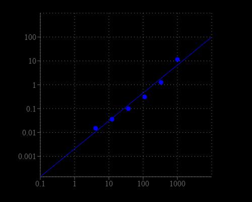
Figure 1. ALT dose response was measured with Amplite Colorimetric Alanine Aminotransferase Assay Kit in a 96-well white, clear-bottom plate using a SpectraMax microplate reader (Molecular Devices).

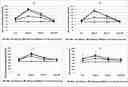
Figure 2. Analysis of serum transaminases following injection with viruses.
(A) ALT concentration following injection with AdEasy, AdEasy-HMGA-6 or PBS as vehicle control into the liver. (B) ALT concentration following injection with AdEasy, AdEasy-HMGA-6 or PBS as vehicle control into the pancreas. (C) AST concentration following injection with AdEasy, AdEasy-HMGA-6 or PBS as vehicle control into the liver. (D) AST concentration following injection with AdEasy, AdEasy-HMGA-6 or PBS as vehicle control into the pancreas. Viruses were injected with a dose of 1.0X10⁸ virus particles / kg of body weight. 20μL of PBS was injected as a vehicle control. The sham control group did not undergo any surgical procedure. Serum samples were collected 6h, 3, 7 or 30 days post-injection of viruses or PBS. All data represent the means ± SD of three mice. Source: A mouse model study of toxicity and biodistribution of a replication defective adenovirus serotype 5 virus with its genome engineered to contain a decoy hyper binding site to sequester and suppress oncogenic HMGA1 as a new cancer treatment therapy, by Faizule Hassan et al., PLOS, Feb. 2018
(A) ALT concentration following injection with AdEasy, AdEasy-HMGA-6 or PBS as vehicle control into the liver. (B) ALT concentration following injection with AdEasy, AdEasy-HMGA-6 or PBS as vehicle control into the pancreas. (C) AST concentration following injection with AdEasy, AdEasy-HMGA-6 or PBS as vehicle control into the liver. (D) AST concentration following injection with AdEasy, AdEasy-HMGA-6 or PBS as vehicle control into the pancreas. Viruses were injected with a dose of 1.0X10⁸ virus particles / kg of body weight. 20μL of PBS was injected as a vehicle control. The sham control group did not undergo any surgical procedure. Serum samples were collected 6h, 3, 7 or 30 days post-injection of viruses or PBS. All data represent the means ± SD of three mice. Source: A mouse model study of toxicity and biodistribution of a replication defective adenovirus serotype 5 virus with its genome engineered to contain a decoy hyper binding site to sequester and suppress oncogenic HMGA1 as a new cancer treatment therapy, by Faizule Hassan et al., PLOS, Feb. 2018
Citations
View all 9 citations: Citation Explorer
Reparative Efficacy of Liposome-Encapsulated Oleanolic Acid against Liver Inflammation Induced by Fine Ambient Particulate Matter and Alcohol in Mice
Authors: Wei, Ching-Ting and Wang, Yu-Wen and Wu, Yu-Chiuan and Lin, Li-Wei and Chen, Chia-Chi and Chen, Chun-Yin and Kuo, Shyh-Ming
Journal: Pharmaceutics (2022): 1108
Authors: Wei, Ching-Ting and Wang, Yu-Wen and Wu, Yu-Chiuan and Lin, Li-Wei and Chen, Chia-Chi and Chen, Chun-Yin and Kuo, Shyh-Ming
Journal: Pharmaceutics (2022): 1108
Pharmacologic IRE1/XBP1s activation promotes systemic adaptive remodeling in obesity
Authors: Madhavan, Aparajita and Kok, Bernard P and Rius, Bibiana and Grandjean, Julia and Alabi, Adekunle and Albert, Verena and Sukiasyan, Ara and Powers, Evan T and Galmozzi, Andrea and Saez, Enrique and others,
Journal: Nature communications (2022): 1--9
Authors: Madhavan, Aparajita and Kok, Bernard P and Rius, Bibiana and Grandjean, Julia and Alabi, Adekunle and Albert, Verena and Sukiasyan, Ara and Powers, Evan T and Galmozzi, Andrea and Saez, Enrique and others,
Journal: Nature communications (2022): 1--9
Decreased autophagosome biogenesis, reduced NRF2, and enhanced ferroptotic cell death are underlying molecular mechanisms of non-alcoholic fatty liver disease
Authors: Liu, Pengfei and Anandhan, Annadurai and Chen, Jingjing and Shakya, Aryatara and Dodson, Matthew and Ooi, Aikseng and Chapman, Eli and White, Eileen and Garcia, Joe GN and Zhang, Donna D
Journal: Redox Biology (2022): 102570
Authors: Liu, Pengfei and Anandhan, Annadurai and Chen, Jingjing and Shakya, Aryatara and Dodson, Matthew and Ooi, Aikseng and Chapman, Eli and White, Eileen and Garcia, Joe GN and Zhang, Donna D
Journal: Redox Biology (2022): 102570
Reparative and toxicity-reducing effects of liposome-encapsulated saikosaponin in mice with liver fibrosis
Authors: Shiu, Li-Yen and Huang, Han Hsiang and Chen, Chun Yin and Cheng, Hsia-Ying and Chen, Chih I and Kuo, Shyh Ming
Journal: Bioscience Reports (2020)
Authors: Shiu, Li-Yen and Huang, Han Hsiang and Chen, Chun Yin and Cheng, Hsia-Ying and Chen, Chih I and Kuo, Shyh Ming
Journal: Bioscience Reports (2020)
Spermidine confers liver protection by enhancing NRF 2 signaling through a MAP 1S-mediated non-canonical mechanism
Authors: Liu, Pengfei and de la Vega, Montserrat Rojo and Dodson, Matthew and Yue, Fei and Shi, Boyun and Fang, Deyu and Chapman, Eli and Liu, Leyuan and Zhang, Donna D
Journal: Hepatology (2019)
Authors: Liu, Pengfei and de la Vega, Montserrat Rojo and Dodson, Matthew and Yue, Fei and Shi, Boyun and Fang, Deyu and Chapman, Eli and Liu, Leyuan and Zhang, Donna D
Journal: Hepatology (2019)
Safety of nanoparticles based on albumin-polymer conjugates as a carrier of nucleotides for pancreatic cancer therapy
Authors: Taguchi, Kazuaki and Lu, Hongxu and Jiang, Yanyan and Hung, Tzong-Tyng and Stenzel, Martina Heide
Journal: Journal of Materials Chemistry B (2018)
Authors: Taguchi, Kazuaki and Lu, Hongxu and Jiang, Yanyan and Hung, Tzong-Tyng and Stenzel, Martina Heide
Journal: Journal of Materials Chemistry B (2018)
Reparative effects of astaxanthin-hyaluronan nanoaggregates against retrorsine-CCl4-induced liver fibrosis and necrosis
Authors: Wu, Yi Jhen and Wu, Yu Chiuan and Chen, I-Fen and Wu, Yi-Lung and Chuang, Chin Wen and Huang, Han Hsiang and Kuo, Shyh Ming
Journal: Molecules (2018): 726
Authors: Wu, Yi Jhen and Wu, Yu Chiuan and Chen, I-Fen and Wu, Yi-Lung and Chuang, Chin Wen and Huang, Han Hsiang and Kuo, Shyh Ming
Journal: Molecules (2018): 726
A mouse model study of toxicity and biodistribution of a replication defective adenovirus serotype 5 virus with its genome engineered to contain a decoy hyper binding site to sequester and suppress oncogenic HMGA1 as a new cancer treatment therapy
Authors: Hassan, Faizule and Lossie, Sarah L and Kasik, Ellen P and Channon, Audrey M and Ni, Shuisong and Kennedy, Michael A
Journal: PloS one (2018): e0192882
Authors: Hassan, Faizule and Lossie, Sarah L and Kasik, Ellen P and Channon, Audrey M and Ni, Shuisong and Kennedy, Michael A
Journal: PloS one (2018): e0192882
References
View all 226 references: Citation Explorer
Pegylated interferon alpha-2b plus ribavirin for Japanese chronic hepatitis C patients with normal alanine aminotransferase
Authors: Kainuma M, Furusyo N, Azuma K, Kajiwara E, Takahashi K, Nomura H, Tanabe Y, Satoh T, Maruyama T, Nakamuta M, Kotoh K, Shimoda S, Hayashi J.
Journal: Hepatol Res (2012): 33
Authors: Kainuma M, Furusyo N, Azuma K, Kajiwara E, Takahashi K, Nomura H, Tanabe Y, Satoh T, Maruyama T, Nakamuta M, Kotoh K, Shimoda S, Hayashi J.
Journal: Hepatol Res (2012): 33
Coexistence of non-alcoholic fatty liver disease with elevated alanine aminotransferase is associated with insulin resistance in young Han males
Authors: Wang R, Lu Q, Feng J, Yin F, Qin C, Liu B, Liu Y, Liu X.
Journal: Endocrine (2012): 70
Authors: Wang R, Lu Q, Feng J, Yin F, Qin C, Liu B, Liu Y, Liu X.
Journal: Endocrine (2012): 70
Levels of Alanine Aminotransferase Confound Use of Transient Elastography to Diagnose Fibrosis in Patients with Chronic HCV Infection
Authors: Tapper EB, Cohen EB, Patel K, Bacon B, Gordon S, Lawitz E, Nelson D, Nasser IA, Challies T, Afdhal N.
Journal: Clin Gastroenterol Hepatol. (2012)
Authors: Tapper EB, Cohen EB, Patel K, Bacon B, Gordon S, Lawitz E, Nelson D, Nasser IA, Challies T, Afdhal N.
Journal: Clin Gastroenterol Hepatol. (2012)
Transient elastographic evaluation in adult subjects without overt liver disease: influence of alanine aminotransferase levels
Authors: Kumar M, Sharma P, Garg H, Kumar R, Bhatia V, Sarin SK.
Journal: J Gastroenterol Hepatol (2011): 1318
Authors: Kumar M, Sharma P, Garg H, Kumar R, Bhatia V, Sarin SK.
Journal: J Gastroenterol Hepatol (2011): 1318
The cross-sectional association between insulin resistance and circulating complement C3 is partly explained by plasma alanine aminotransferase, independent of central obesity and general inflammation (the CODAM study)
Authors: van Greevenbroek MM, Jacobs M, van der Kallen CJ, Vermeulen VM, Jansen EH, Schalkwijk CG, Ferreira I, Feskens EJ, Stehouwer CD.
Journal: Eur J Clin Invest (2011): 372
Authors: van Greevenbroek MM, Jacobs M, van der Kallen CJ, Vermeulen VM, Jansen EH, Schalkwijk CG, Ferreira I, Feskens EJ, Stehouwer CD.
Journal: Eur J Clin Invest (2011): 372
Nonalcoholic fatty liver disease is associated with blood pressure in hypertensive and nonhypertensive individuals from the general population with normal levels of alanine aminotransferase
Authors: Lopez-Suarez A, Guerrero JM, Elvira-Gonzalez J, Beltran-Robles M, Canas-Hormigo F, Bascunana-Quirell A.
Journal: Eur J Gastroenterol Hepatol (2011): 1011
Authors: Lopez-Suarez A, Guerrero JM, Elvira-Gonzalez J, Beltran-Robles M, Canas-Hormigo F, Bascunana-Quirell A.
Journal: Eur J Gastroenterol Hepatol (2011): 1011
Ferritin/alanine aminotransferase ratio as a possible marker for predicting the prognosis of acute liver injury
Authors: Ozawa E, Abiru S, Nagaoka S, Yano K, Komori A, Migita K, Yatsuhashi H, Taura N, Ichikawa T, Ishibashi H, Nakao K.
Journal: J Gastroenterol Hepatol (2011): 1326
Authors: Ozawa E, Abiru S, Nagaoka S, Yano K, Komori A, Migita K, Yatsuhashi H, Taura N, Ichikawa T, Ishibashi H, Nakao K.
Journal: J Gastroenterol Hepatol (2011): 1326
Changes in serum levels of HBV DNA and alanine aminotransferase determine risk for hepatocellular carcinoma
Authors: Chen CF, Lee WC, Yang HI, Chang HC, Jen CL, Iloeje UH, Su J, Hsiao CK, Wang LY, You SL, Lu SN, Chen CJ.
Journal: Gastroenterology (2011): 1240
Authors: Chen CF, Lee WC, Yang HI, Chang HC, Jen CL, Iloeje UH, Su J, Hsiao CK, Wang LY, You SL, Lu SN, Chen CJ.
Journal: Gastroenterology (2011): 1240
Association of serum alanine aminotransferase and gamma-glutamyltransferase levels within the reference range with metabolic syndrome and nonalcoholic fatty liver disease
Authors: Oh HJ, Kim TH, Sohn YW, Kim YS, Oh YR, Cho EY, Shim SY, Shin SR, Han AL, Yoon SJ, Kim HC.
Journal: Korean J Hepatol (2011): 27
Authors: Oh HJ, Kim TH, Sohn YW, Kim YS, Oh YR, Cho EY, Shim SY, Shin SR, Han AL, Yoon SJ, Kim HC.
Journal: Korean J Hepatol (2011): 27
Non-alcoholic fatty liver disease's prevalence and impact on alanine aminotransferase associated with metabolic syndrome in the Chinese
Authors: Hou XH, Zhu YX, Lu HJ, Chen HF, Li Q, Jiang S, Xiang KS, Jia WP.
Journal: J Gastroenterol Hepatol (2011): 722
Authors: Hou XH, Zhu YX, Lu HJ, Chen HF, Li Q, Jiang S, Xiang KS, Jia WP.
Journal: J Gastroenterol Hepatol (2011): 722
Application notes
Restriction of Advanced Glycation End Products Improves Insulin Resistance in Human Type 2 Diabetes
Design of potent inhibitors of acetylcholinesterase using morin as the starting compound
Acetylcholinesterase Inhibitory Activity of Pigment Echinochrome A
Induction of Neurite Outgrowth in PC12 Cells
Induction of Neuritogenesis in PC12 Cells by a Pulsed Electromagnetic Field
Design of potent inhibitors of acetylcholinesterase using morin as the starting compound
Acetylcholinesterase Inhibitory Activity of Pigment Echinochrome A
Induction of Neurite Outgrowth in PC12 Cells
Induction of Neuritogenesis in PC12 Cells by a Pulsed Electromagnetic Field
FAQ
Why should I use an absorbance ratio at A575nm/A605nm when using most of your Amplite® Colorimetric Assay Kits?
How should I reconstitute an NADPH standard?
Will Amplite® Fluorimetric NAD/NADH Ratio Assay Kit *Red Fluorescence* work with NADP/NADPH? Can this kit measure NADP+ and NADPH?
What is the concentration of calcium inside cells?
What assay kits measure NADP/NADPH from cell samples?
How should I reconstitute an NADPH standard?
Will Amplite® Fluorimetric NAD/NADH Ratio Assay Kit *Red Fluorescence* work with NADP/NADPH? Can this kit measure NADP+ and NADPH?
What is the concentration of calcium inside cells?
What assay kits measure NADP/NADPH from cell samples?