Cell Signaling
In biology, cell signaling is a network of communications that governs basic and complex cellular processes. Depending upon the nature of the signal, it can be classified as either biochemical or mechanical. Biochemical signals are generated by biochemical molecules such as proteins, lipids, ions or hormones. Detection of these biochemical agents may shed light on the state of that particular type of signaling pathway.
Disruption of signaling networks often leads to diseases such as cancer, immune diseases and metabolic disorders. By understanding signaling pathways, researchers and clinicians may be able to treat disease conditions effectively and efficiently. AAT Bioquest offers a broad range of assays to conduct cost-effective analysis of various enzymes, proteins or small molecules involved cell signaling pathways. Measure activities such as phosphorylation, methylation and acetylation with unparalleled sensitivity and reproducibility.
Disruption of signaling networks often leads to diseases such as cancer, immune diseases and metabolic disorders. By understanding signaling pathways, researchers and clinicians may be able to treat disease conditions effectively and efficiently. AAT Bioquest offers a broad range of assays to conduct cost-effective analysis of various enzymes, proteins or small molecules involved cell signaling pathways. Measure activities such as phosphorylation, methylation and acetylation with unparalleled sensitivity and reproducibility.
Adenosine Diphosphate Assays

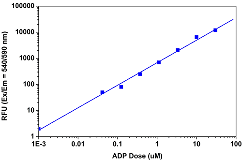
ADP dose response was measured with the PhosphoWorks™ Fluorimetric ADP Assay Kit (Cat NO. 21655) in a solid black 384-well plate using a Gemini fluorescence microplate reader (Molecular Devices). As low as 0.3 µM ADP can be detected with 15, 30 minutes and 1 hour incubation (Z' factor =0.65).
The PhosphoWorks™ Fluorimetric ADP assay offers a sensitive method for monitoring ADP formation. In this enzyme-coupled assay, ADP formation is measured by monitoring kinase activity due to their proportional relationship.
Table 1. Biochemical assays for measuring ADP activity, formation or depletion.
| Assay ▲ ▼ | Ex (nm) ▲ ▼ | Em (nm) ▲ ▼ | Cutoff (nm)¹ ▲ ▼ | Microplate Type ▲ ▼ | Unit Size ▲ ▼ | Cat No. ▲ ▼ |
| PhosphoWorks™ Fluorimetric ADP Assay Kit *Red Fluorescence* | 540 | 590 | 570 | Solid black | 100 tests | 21655 |
- Fluorescence microplate instrument specifications.
Adenosine Triphosphate Assays

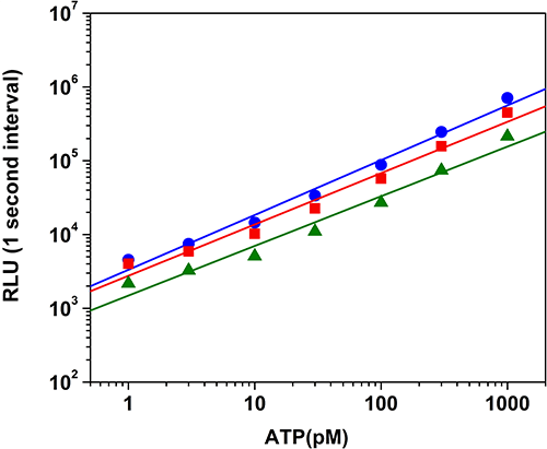
ATP dose response was measured with the PhosphoWorks™ Luminescence ATP Assay Kit (Cat No. 21610) on a 96-well white plate using a NOVOstar plate reader (BMG Labtech). The kit can detect 3 pmol ATP with 2 hours incubation (Z' factor = 0.6, blue 30 minutes, red 1 hour, and green 2 hours). The integration time was 1 sec. The half life is more than 1.5 hours.
Our PhosphoWorks™ Luminometric ATP assays provide a fast, simple and homogenous bioluminescence method that can be used for the determination of cell viability, cytotoxicity and proliferation in mammalian cells by ATP detection.
Table 2. Biochemical assays for measuring ATP activity, formation or depletion.
| Assay ▲ ▼ | Ex/Abs (nm) ▲ ▼ | Em (nm) ▲ ▼ | Cutoff (nm)¹ ▲ ▼ | Microplate Type ▲ ▼ | Unit Size ▲ ▼ | Cat No. ▲ ▼ |
| PhosphoWorks™ Fluorimetric ATP Assay Kit | 540 | 590 | 570 | Solid black | 100 tests | 21620 |
| PhosphoWorks™ Colorimetric ATP Assay Kit | 570 | - | - | Clear bottom | 100 tests | 21617 |
| PhosphoWorks™ Luminometric ATP Assay Kit *Maximized Luminescence* | - | - | - | Solid white | 1 plate | 21610 |
| PhosphoWorks™ Luminometric ATP Assay Kit *Maximized Luminescence* | - | - | - | Solid white | 10 plate | 21621 |
| Cell Meter™ Live Cell ATP Assay Kit | - | - | - | Black wall/clear bottom | 100 Tests | 23015 |
- Fluorescence microplate instrument specifications.
Cyclic Adenosine Monophosphate Assays

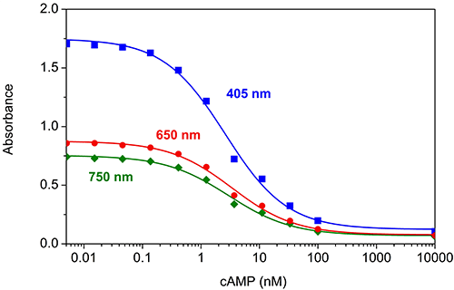
cAMP dose response was measured with Screen Quest™ Colorimetric ELISA cAMP Assay Kit (Cat#36370) in a clear 96-well plate with a SpectraMax microplate reader. The Absorbance can be read at 405 nm (blue line), 650 nm (red line) or 740 nm (Green line), the data in figure B are from the incubation with Amplite™ Green for 3 hours.
Table 3. Biochemical assays for monitoring the activation of adenylyl cyclase in G-protein coupled receptor systems.
| Assay ▲ ▼ | Ex/Abs (nm) ▲ ▼ | Em (nm) ▲ ▼ | Cutoff (nm)¹ ▲ ▼ | Microplate Type ▲ ▼ | Unit Size ▲ ▼ | Cat No. ▲ ▼ |
| Screen Quest™ Fluorimetric ELISA cAMP Assay Kit | 540 | 590 | 570 | Solid black plate | 1 plates | 36373 |
| Screen Quest™ Fluorimetric ELISA cAMP Assay Kit | 540 | 590 | 570 | Solid black plate | 10 plates | 36374 |
| Screen Quest™ Colorimetric ELISA cAMP Assay Kit | 405, 650, 740 | - | - | Clear plate | 1 plate | 36370 |
| Screen Quest™ Colorimetric ELISA cAMP Assay Kit | 405, 650, 740 | - | - | Clear plate | 10 plates | 36371 |
| Screen Quest™ FRET No Wash cAMP Assay Kit | - | - | - | Solid black plate, black wall/clear bottom plate | 1 plates | 36379 |
| Screen Quest™ FRET No Wash cAMP Assay Kit | - | - | - | Solid black plate, black wall/clear bottom plate | 10 plates | 36380 |
| Screen Quest™ FRET No Wash cAMP Assay Kit | - | - | - | Solid black plate, black wall/clear bottom plate | 50 plates | 36381 |
Table 4. Probes For Detecting And Monitoring cGAMP Activity
| Cat# ▲ ▼ | Product Name ▲ ▼ | Unit Size ▲ ▼ |
| 20310 | 2',3'-cGAMP | 100 µg |
| 20311 | 2',3'-cGAMP | 1 mg |
| 20313 | 2',3'-cGAMP, sodium salt | 100 µg |
| 20316 | 2',3'-cGAMP-Biotin conjugate | 100 µg |
| 20318 | 2',3'-cGAMP-Cy5 conjugate | 100 µg |
| 20320 | 2’,3’-cGAMP-iFluor 488 conjugate | 100 µg |
| 20322 | 2’,3’-cGAMP-iFluor 647 conjugate | 100 µg |
| 20330 | 2’,3’-cGAMP-DBCO conjugate | 100 µg |
| 20332 | 2’,3’-cGAMP azide | 100 µg |
| 20334 | 2’,3’-cGAMP succinimidyl ester | 100 µg |