logo
AAT Bioquest

StrandBrite™ Green Fluorimetric RNA Quantitation Kit *High Selectivity*

RNA dose response measured with StrandBrite™ Green Fluorimetric RNA Quantitation Kit (Cat#17657) in a solid black 96-well microplate using a Gemini microplate reader (Molecular Devices). 
Ordering information
Price
Catalog Number
Unit Size
Quantity
Add to cart
Additional ordering information
Telephone1-800-990-8053
Fax1-800-609-2943
Emailsales@aatbio.com
InternationalSee distributors
Bulk requestInquire
Custom sizeInquire
ShippingStandard overnight for United States, inquire for international
Request quotation
Spectral properties
Excitation (nm)509
Emission (nm)527
Storage, safety and handling
H-phraseH303, H313, H340
Hazard symbolT
Intended useResearch Use Only (RUO)
R-phraseR20, R21, R68
UNSPSC41116134

OverviewpdfSDSpdfProtocol


Excitation (nm)
509
Emission (nm)
527
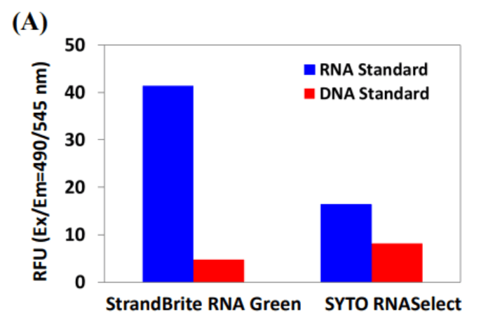
The major challenge to analyze RNA in live cells is the interferences caused by DNA. To address these difficulties, AAT Bioquest has developed the StrandBrite™ RNA Green, an excellent RNA-selective probe that generates significantly enhanced green fluorescence upon binding to RNA. It has been successfully used for flow cytometric analysis of live cells. StrandBrite™ RNA Green readily gets into live cells. It has the excitation/emission of 490/540 nm. In the DNase digest test, no significant change of fluorescence intensity in fixed cells stained with StrandBrite RNA Green was observed. In contrast, after RNase digestion, the initial fluorescence signal decreased immediately. These results indicate that initial fluorescence signal was generated from the specific interaction of StrandBrite RNA Green with RNA in cells. Short exposure of live cells to antinomycin D did cause inhibition of RNA synthesis during 6 hours after drug removal in a dose-dependent manner. These data demonstrate that StrandBrite RNA Green is a sensitive RNA-selective dye for staining nucleolar RNA in live and fixed cells. StrandBrite RNA Green has less DNA interferences than the commonly used SYTO® RNASelect™ dye. StrandBrite™ RNA Green is a highly RNA-selective fluorescent probe. Due to its excellent cell permeability and spectral properties, it has been successfully used for flow cytometric RNA analysis and fluorescence microscope in live cells. It can be well excited with the 488 nm blue laser and monitored in FITC channel. StrandBrite™ RNA Green provides a valuable method for identifying and labeling cells with a single incubation step and can discriminate RNA from DNA with better selectivity than the commonly used SYTO® RNASelect™.

Platform


Fluorescence microplate reader

Excitation490 nm
Emission540 nm
Cutoff515 nm
Recommended plateSolid black

Components


Example protocol


AT A GLANCE

Protocol Summary
  1. Prepare StrandBrite™ RNA Green working solution (100 µL)
  2. Add RNA standards or test samples (100 µL)
  3. Incubate at room temperature for 2 - 5 minutes
  4. Monitor the fluorescence intensity at Ex/Em = 490/540 nm (Cutoff = 515 nm) 
Important      The following protocol is an example for quantifying RNA with StrandBrite™ Green Fluorimetric RNA Quantitation Kit. Allow all the components to warm to room temperature before opening. To prevent RNase contamination of the StrandBrite™ reagent and kit components, always use clean disposable gloves while handling all materials. Use nucleasefree water, and sterile, disposable polypropylene plastic ware for reagent preparation. No data are available addressing the mutagenicity or toxicity of StrandBrite™ RNA Green. Because this reagent binds to nucleic acids, it should be treated as a potential mutagen and handled with appropriate care. The DMSO stock solution should be handled with particular caution.

PREPARATION OF STOCK SOLUTIONS

Unless otherwise noted, all unused stock solutions should be divided into single-use aliquots and stored at -20 °C after preparation. Avoid repeated freeze-thaw cycles.

Assay buffer (1X)
Dilute 10X Assay Buffer (Component B) in sterile, distilled, nuclease-free water to make 1X Assay Buffer.

PREPARATION OF STANDARD SOLUTION

For convenience, use the Serial Dilution Planner:
https://www.aatbio.com/tools/serial-dilution/17657


RNA standard
Add 10 µL of 2 mg/mL RNA Standard (Component C) to 990 µL of 1X Assay buffer to make 20 µg/mL RNA standard solution (RS7). Take 20 µg/mL RNA standard solution (RS7) and perform 1:2 serial dilutions in 1X Assay buffer to get serially diluted RNA standards (RS6 - RS1).

PREPARATION OF WORKING SOLUTION

Add 10 μL of StrandBrite™ RNA Green (Component A) into 1.99 mL of 1X Assay buffer to make StrandBrite™ RNA Green working solution. Protect StrandBrite™ RNA Green working solution from light by covering it with foil or placing it in the dark.
Note     We recommend preparing this solution in a plastic container rather than glass, as the dye may adsorb to glass surfaces. For best results, this solution should be used within a few hours of its preparation.

SAMPLE EXPERIMENTAL PROTOCOL

Table 1. Layout of RNA standards and test samples in a solid black 96-well microplate. RS=RNA Standards (RS1 - RS7, 0.313 to 20 µg/mL); BL=Blank Control; TS=Test Samples.
BLBLTSTS
RS1RS1......
RS2RS2......
RS3RS3
RS4RS4
RS5RS5
RS6RS6
RS7RS7
Table 2. Reagent composition for each well.
WellVolumeReagent
RS1 - RS7100 µLSerial Dilutions (0.313 to 20 µg/mL)
BL100 µL1X Assay Buffer
TS100 µLtest sample
  1. Prepare RNA standards (RS), blank controls (BL), and test samples (TS) according to the layout provided in Tables 1 and 2. For a 384-well plate, use 25 µL of reagent per well instead of 50 µL.
  2. Add 100 µL of StrandBrite™ RNA Green working solution to each well of RNA standard, blank control, and test samples to make the total RNA assay volume of 200 µL/well. For a 384-well plate, add 25 µL of StrandBrite™ RNA Green working solution into each well instead, for a total volume of 50 µL/well.
  3. Incubate the reaction at room temperature for 2 to 5 minutes, protected from light.
  4. Monitor the fluorescence increase with a fluorescence microplate reader at Ex/Em = 490/540 nm (Cutoff = 515 nm).
    Note     To minimize photobleaching effects, keep the time for fluorescence measurement constant for all samples. 

Spectrum


Open in Advanced Spectrum Viewer
spectrum

Spectral properties

Excitation (nm)509
Emission (nm)527

Images


Citations


View all 3 citations: Citation Explorer
The nucleolar shell provides anchoring sites for DNA untwisting
Authors: Fukute, Jumpei and Maki, Koichiro and Adachi, Taiji
Journal: Communications Biology (2024): 83
Anticancer Activity of Reconstituted Ribonuclease S-Decorated Artificial Viral Capsid
Authors: Liang, Yingbing and Furukawa, Hiroto and Sakamoto, Kentarou and Inaba, Hiroshi and Matsuura, Kazunori
Journal: ChemBioChem (2022): e202200220
A nanoscale metal organic frameworks-based vaccine synergises with PD-1 blockade to potentiate anti-tumour immunity
Authors: Li, Xia and Wang, Xiupeng and Ito, Atsuo and Tsuji, Noriko M
Journal: Nature communications (2020): 1--15

References


View all 31 references: Citation Explorer
Inhibitors of Streptococcus pneumoniae surface endonuclease EndA discovered by high-throughput screening using a PicoGreen fluorescence assay
Authors: Peterson EJ, Kireev D, Moon AF, Midon M, Janzen WP, Pingoud A, Pedersen LC, Singleton SF.
Journal: J Biomol Screen (2013): 247
Validation of a PicoGreen-based DNA quantification integrated in an RNA extraction method for two-dimensional and three-dimensional cell cultures
Authors: Chen Y, Sonnaert M, Roberts SJ, Luyten FP, Schrooten J.
Journal: Tissue Eng Part C Methods (2012): 444
Characterization of PicoGreen interaction with dsDNA and the origin of its fluorescence enhancement upon binding
Authors: Dragan AI, Casas-Finet JR, Bishop ES, Strouse RJ, Schenerman MA, Geddes CD.
Journal: Biophys J (2010): 3010
Comparison of SYBR Green I-, PicoGreen-, and [3H]-hypoxanthine-based assays for in vitro antimalarial screening of plants from Nigerian ethnomedicine
Authors: Abiodun OO, Gbotosho GO, Ajaiyeoba EO, Happi CT, Hofer S, Wittlin S, Sowunmi A, Brun R, Oduola AM.
Journal: Parasitol Res (2010): 933
Metal-enhanced PicoGreen fluorescence: application to fast and ultra-sensitive pg/ml DNA quantitation
Authors: Dragan AI, Bishop ES, Casas-Finet JR, Strouse RJ, Schenerman MA, Geddes CD.
Journal: J Immunol Methods (2010): 95
Quantification of dsDNA using the Hitachi F-7000 Fluorescence Spectrophotometer and PicoGreen dye
Authors: Moreno LA, Cox KL.
Journal: J Vis Exp. (2010)
Factors affecting quantification of total DNA by UV spectroscopy and PicoGreen fluorescence
Authors: Holden MJ, Haynes RJ, Rabb SA, Satija N, Yang K, Blasic JR, Jr.
Journal: J Agric Food Chem (2009): 7221
Development and characterization of a novel host cell DNA assay using ultra-sensitive fluorescent nucleic acid stain "PicoGreen"
Authors: Ikeda Y, Iwakiri S, Yoshimori T.
Journal: J Pharm Biomed Anal (2009): 997
Enhanced DNA dynamics due to cationic reagents, topological states of dsDNA and high mobility group box 1 as probed by PicoGreen
Authors: Noothi SK, Kombrabail M, Kundu TK, Krishnamoorthy G, Rao BJ.
Journal: FEBS J (2009): 541
Label-free DNA sequence detection with enhanced sensitivity and selectivity using cationic conjugated polymers and PicoGreen
Authors: Ren X, Xu QH.
Journal: Langmuir (2009): 43