Products
Technologies
Applications
Services
Resources
Selection Guides
About
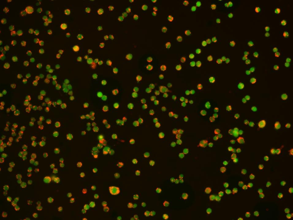
Protonex™ Red 600-Latex Bead Conjugate
Protonex™ Red-latex bead conjugate demonstrated pH-dependent fluorescence. Unlike most of the existing fluorescent dyes that are more fluorescent at higher pH, acidic conditions enhance the fluorescence of Protonex™ Red-latex bead conjugate. The fluorescence of Protonex™ Red-latex bead conjugate dramatically increases as pH decreases from neutral to the acidic, making it a robust tool to study phagocytosis and its regulation by drugs and/or environmental factors. The lack of fluorescence outside the cell eliminates the wash steps. Protonex™ Red-latex bead conjugate provides a powerful tool to study phagocytosis. Protonex™ Red-latex bead conjugate is low fluorescent outside the cells, but fluoresce brightly red in acidic compartments (such as phagosomes, lysosomes and endosomes). This Protonex™ Red-latex bead conjugate can be also used for multiplexing cell functional analysis with green dyes such as GFP, Fluo-8®, calcein, or FITC-labeled antibodies. Protonex™ Red has the spectral properties similar to those of Texas Red, making the common filter set of Texas Red readily available to the assays of Protonex™ Red.
| Catalog | Size | Price | Quantity |
|---|---|---|---|
| 21209 | 1 mL | Price |

Spectral properties
| Excitation (nm) | 576 |
| Emission (nm) | 597 |
Storage, safety and handling
| H-phrase | H303, H313, H333 |
| Hazard symbol | XN |
| Intended use | Research Use Only (RUO) |
| R-phrase | R20, R21, R22 |
| Storage | Refrigerated (2-8 °C); Minimize light exposure |
| UNSPSC | 12352200 |
Instrument settings
| Fluorescence microscope | |
| Excitation | Texas Red filter set |
| Emission | Texas Red filter set |
| Recommended plate | Black wall/clear bottom |
Documents
Contact us
| Telephone | |
| Fax | |
| sales@aatbio.com | |
| International | See distributors |
| Bulk request | Inquire |
| Custom size | Inquire |
| Technical Support | Contact us |
| Request quotation | Request |
| Purchase order | Send to sales@aatbio.com |
| Shipping | Standard overnight for United States, inquire for international |
Page updated on December 9, 2025