logo
AAT Bioquest

Cell Meter™ Fluorimetric Intracellular Total ROS Activity Assay Kit*Green Fluorescence*

Fluorescence images of simultaneous detection of intracellular nitric oxide (NO) and total ROS in RAW 264.7 macrophage. Cells were co-stained with Nitrixyte™ Orange (Red) and Amplite® ROS Green (Green). The cells were then treated with or without 20 µg/mL of lipopolysaccharide (LPS), 1 mM L-arginine (L-Arg) and 50 µM Pyocyanin (Pyo) at 37°C for 16 hours. The fluorescence signals were measured using fluorescence microscope equipped with TRITC (Nitrixyte™ Orange, Red) and FITC (Amplite® ROS Green, Green) filter sets, simultaneously.
Detection of ROS in Jurkat cells with Cell Meter&trade; Fluorimetric Intracellular Total ROS Activity Assay Kit. Jurkat cells were seeded on the same day at 300,000 cells/100&micro;L/well in a Costar black wall/clear bottom 96-well plate. The ROS assay loading solution (100 &micro;L/well) was added and incubated in a 5% CO2, 37 &deg;C incubator for 1 hour. And then the cells were treated with 1mM, 0.1mM H<sub>2</sub>O<sub>2</sub> or without H<sub>2</sub>O<sub>2</sub> for 30 minutes. The fluorescence signal was monitored at Ex/Em = 490/525 nm (cutoff at 515 nm) with bottom read mode using FlexStation (Molecular Devices).
Ordering information
Price
Catalog Number
Unit Size
Quantity
Add to cart
Additional ordering information
Telephone1-800-990-8053
Fax1-800-609-2943
Emailsales@aatbio.com
InternationalSee distributors
Bulk requestInquire
Custom sizeInquire
ShippingStandard overnight for United States, inquire for international
Request quotation
Storage, safety and handling
H-phraseH303, H313, H333
Hazard symbolXN
Intended useResearch Use Only (RUO)
R-phraseR20, R21, R22
UNSPSC12352200
Related products
Cell Meter™ Intracellular Fluorimetric Hydrogen Peroxide Assay Kit *Green Fluorescence*
Cell Meter™ Intracellular Fluorimetric Hydrogen Peroxide Assay Kit *Blue Fluorescence*
Cell Meter™ Intracellular Fluorimetric Hydrogen Peroxide Assay Kit *Blue Fluorescence Optimized for Flow Cytometry*
Cell Meter™ Intracellular Fluorimetric Hydrogen Peroxide Assay Kit *Green Fluorescence Optimized for Flow Cytometry*
Cell Meter™ Mitochondrial Hydroxyl Radical Detection Kit *Red Fluorescence*
Cell Meter™ Fluorimetric Mitochondrial Superoxide Activity Assay Kit *Green Fluorescence*
Cell Meter™ Fluorimetric Intracellular Peroxynitrite Assay Kit *Green Fluorescence*
Cell Meter™ Fluorimetric Intracellular Peroxynitrite Assay Kit *Optimized for Flow Cytometry*
Cell Meter™ Fluorimetric Intracellular Nitric Oxide (NO) Activity Assay Kit *Orange Fluorescence Optimized for Microplate Reader*
Cell Meter™ Fluorimetric Intracellular Nitric Oxide (NO) Activity Assay Kit *Orange Fluorescence Optimized for Flow Cytometry*
Cell Meter™ Fluorimetric Intracellular Nitric Oxide (NO) Activity Assay Kit *Red Fluorescence Optimized for Flow Cytometry*
Cell Meter™ Live Cell Caspase 3/7 Binding Assay Kit *Green Fluorescence*
Cell Meter™ Live Cell Caspase 3/7 Binding Assay Kit *Red Fluorescence*
Cell Meter™ Live Cell Caspase 1 Binding Assay Kit *Green Fluorescence*
Cell Meter™ Live Cell Caspase 2 Binding Assay Kit *Green Fluorescence*
Cell Meter™ Live Cell Caspase 6 Binding Assay Kit *Green Fluorescence*
Cell Meter™ Live Cell Caspase 8 Binding Assay Kit *Green Fluorescence*
Cell Meter™ Live Cell Caspase 9 Binding Assay Kit *Green Fluorescence*
Cell Meter™ Live Cell Caspase 10 Binding Assay Kit *Green Fluorescence*
Cell Meter™ Live Cell Caspase 13 Binding Assay Kit *Green Fluorescence*
Cell Meter™ No-Wash Live Cell Caspase 3/7 Activity Assay Kit *Blue Fluorescence*
Cell Meter™ No-Wash Live Cell Caspase 3/7 Activity Assay Kit *Red Fluorescence*
Cell Meter™ Fluorimetric Intracellular pH Assay Kit
Cell Meter™ Fluorimetric Cell Cytotoxicity Assay Kit
Cell Meter™ Cell Viability Assay Kit *Red Fluorescence*
Cell Meter™ Cell Viability Assay Kit *Blue Fluorescence with 405 nm Excitation*
Cell Meter™ Cell Viability Assay Kit *Blue Fluorescence*
Cell Meter™ Cell Viability Assay Kit *Green Fluorescence*
Cell Meter™ Cell Viability Assay Kit *NIR Fluorescence Optimized for Fluorescence Microplate Reader*
Cell Meter™ Phosphatidylserine Apoptosis Assay Kit *Blue Fluorescence Optimized for Microplate Readers*
Cell Meter™ Phosphatidylserine Apoptosis Assay Kit *Green Fluorescence Optimized for Microplate Readers*
Cell Meter™ Phosphatidylserine Apoptosis Assay Kit *Red Fluorescence Optimized for Microplate Readers*
Cell Meter™ Phosphatidylserine Apoptosis Assay Kit *Deep Red Fluorescence Optimized for Microplate Readers*
Cell Meter™ Phosphatidylserine Apoptosis Assay Kit *Orange Fluorescence Optimized for Microplate Readers*
Cell Meter™ Caspase 3/7 Activity Apoptosis Assay Kit *Blue Fluorescence*
Cell Meter™ Caspase 3/7 Activity Apoptosis Assay Kit *Green Fluorescence*
Cell Meter™ Caspase 3/7 Activity Apoptosis Assay Kit *Red Fluorescence*
Cell Meter™ Caspase 8 Activity Apoptosis Assay Kit *Green Fluorescence*
Cell Meter™ Caspase 9 Activity Apoptosis Assay Kit *Green Fluorescence*
Cell Meter™ JC-10 Mitochondrion Membrane Potential Assay Kit *Optimized for Microplate Assays*
Cell Meter™ JC-10 Mitochondrion Membrane Potential Assay Kit *Optimized for Flow Cytometry Assays*
Cell Meter™ NIR Mitochondrion Membrane Potential Assay Kit *Optimized for Flow Cytometry*
Cell Meter™ NIR Mitochondrion Membrane Potential Assay Kit *Optimized for Microplate Reader*
Cell Meter™ Mitochondrion Membrane Potential Assay Kit *Orange Fluorescence Optimized for Flow Cytometry*
Cell Meter™ Mitochondrion Membrane Potential Assay Kit *Orange Fluorescence Optimized for Microplate Reader*
Cell Meter™ Intracellular GSH Assay Kit *Optimized for Flow Cytometry*
Cell Meter™ Nuclear Apoptosis Assay Kit *Green Fluorescence Optimized for Flow Cytometry*
Cell Meter™ Caspase 8 Activity Apoptosis Assay Kit *Blue Fluorescence*
Cell Meter™ Caspase 9 Activity Apoptosis Assay Kit *Blue Fluorescence*
Cell Meter™ Caspase 8 Activity Apoptosis Assay Kit *Red Fluorescence*
Cell Meter™ Caspase 9 Activity Apoptosis Assay Kit *Red Fluorescence*
Cell Meter™ Multiplexing Caspase 3/7, 8 and 9 Activity Assay Kit *Triple Fluorescence Colors*
Cell Meter™ Generic Fluorimetric Caspase Activity Assay Kit *Green Fluorescence Optimized for Flow Cytometry*
Cell Meter™ Generic Fluorimetric Caspase Binding Assay Kit *Red Fluorescence Optimized for Flow Cytometry*
Cell Meter™ Caspase 3/7 Activity Apoptosis Assay Kit *Green Fluorescence Optimized for Flow Cytometry*
Cell Meter™ Annexin V Binding Apoptosis Assay Kit *Green Fluorescence Optimized for Flow Cytometry*
Cell Meter™ Annexin V Binding Apoptosis Assay Kit *Orange Fluorescence Optimized for Flow Cytometry*
Cell Meter™ Annexin V Binding Apoptosis Assay Kit *Red Fluorescence Optimized for Flow Cytometry*
Cell Meter™ Annexin V Binding Apoptosis Assay Kit *Deep Red Fluorescence Optimized for Flow Cytometry*
Cell Meter™ Annexin V Binding Apoptosis Assay Kit *Blue Fluorescence Excited at 405 nm*
Cell Meter™ Annexin V Binding Apoptosis Assay Kit *Green Fluorescence Excited at 405 nm*
Cell Meter™ Annexin V Binding Apoptosis Assay Kit *Orange Fluorescence Excited at 405 nm*
Cell Meter™ Phosphatidylserine Apoptosis Assay Kit *Green Fluorescence Optimized for Flow Cytometry*
Cell Meter™ Phosphatidylserine Apoptosis Assay Kit *Deep Red Fluorescence Optimized for Flow Cytometry*
Cell Meter™ Phosphatidylserine Apoptosis Assay Kit *Blue Fluorescence Excited at 405 nm*
Cell Meter™ Phosphatidylserine Apoptosis Assay Kit *Green Fluorescence Excited at 405 nm*
Cell Meter™ APC-Annexin V Binding Apoptosis Assay Kit *Optimized for Flow Cytometry*
Cell Meter™ PE-Annexin V Binding Apoptosis Assay Kit *Optimized for Flow Cytometry*
Cell Meter™ Apoptotic and Necrotic Multiplexing Detection Kit I *Triple Fluorescence Colors*
Cell Meter™ Fluorimetric Live Cell Cycle Assay Kit *Green Fluorescence Optimized for Flow Cytometry*
Cell Meter™ Fluorimetric Fixed Cell Cycle Assay Kit *Red Fluorescence Optimized for Flow Cytometry*
Cell Meter™ Apoptotic and Necrotic Multiplexing Detection Kit II *Triple Fluorescence Colors*
Cell Meter™ Live Cell TUNEL Apoptosis Assay Kit *Red Fluorescence*
Cell Meter™ Fluorimetric Live Cell Cycle Assay Kit *Optimized for 405 nm Violet Laser Excitation*
Cell Meter™ Live Cell Caspase 3/7 and Phosphatidylserine Detection Kit *Triple Fluorescence Colors*
Cell Meter™ Fluorimetric Mitochondrial Superoxide Activity Assay Kit*Optimized for Flow Cytometry*
Cell Meter™ Fluorimetric Mitochondrial Superoxide Activity Assay Kit*Optimized for Microplate Reader*
Cell Meter™ Autophagy Assay Kit *Blue Fluorescence*
Cell Meter™ Autophagy Fluorescence Imaging Kit
Cell Meter™ Autophagy Assay Kit *Green Fluorescence*
Cell Meter™ FITC-Annexin V Binding Apoptosis Assay Kit *Optimized for Flow Cytometry*
Cell Meter™ Fluorimetric Intracellular Nitric Oxide (NO) Activity Assay Kit *NIR Fluorescence Optimized for Microplate Reader*
Cell Meter™ Fluorimetric Intracellular Nitric Oxide (NO) Activity Assay Kit *NIR Fluorescence Optimized for Flow Cytometry*
Cell Meter™ Colorimetric WST-8 Cell Quantification Kit
Cell Meter™ Multiplexing Live, Apoptotic and Necrotic Cell Detection Kit III *Triple Fluorescence Colors*
Cell Meter™ 2-NBDG Glucose Uptake Assay Kit
Cell Meter™ Intracellular NADH/NADPH Fluorescence Imaging Kit *Red Fluorescence*
Cell Meter™ Intracellular NADH/NADPH Flow Cytometric Analysis Kit *Red Fluorescence*
Cell Meter™ Live Cell TUNEL Apoptosis Assay Kit *Green Fluorescence*
Cell Meter™ No Wash and Probenecid-Free Endpoint Calcium Assay Kit *Optimized for microplate reader*
Cell Meter™ Mitochondrion Membrane Potential Assay Kit *Red Fluorescence Optimized for Flow Cytometry*
Cell Meter™ Mitochondrion Membrane Potential Assay Kit *Red Fluorescence Optimized for Microplate Reader*
Cell Meter™ Intracellular Colorimetric Lipid Peroxidation (MDA) Assay Kit
Cell Meter™ Flow Cytometric Calcium Assay Kit
Cell Meter™ Fluorimetric Live Cell Cycle Assay Kit *Red Fluorescence Optimized for Flow Cytometry*
Cell Meter™ Fluorimetric Cellular Lipid Peroxidation Assay Kit
Cell Meter™ Intracellular NADH/NADPH Fluorescence Imaging Kit *Deep Red Fluorescence*
Cell Meter™ Intracellular NADH/NADPH Flow Cytometric Analysis Kit *Deep Red Fluorescence*
Cell Meter™ Intracellular GSH Assay Kit *Optimized for Flow Cytometry with 405 nm excitation*
Cell Meter™ Mitochondrial Autophagy Imaging Kit *Red Fluorescence*
Cell Meter™ BX520 fixable viability dye
Cell Meter™ BX590 fixable viability dye
Cell Meter™ BX650 fixable viability dye
Cell Meter™ IX830 fixable viability dye
Cell Meter™ RX660 fixable viability dye
Cell Meter™ RX700 fixable viability dye
Cell Meter™ RX780 fixable viability dye
Cell Meter™ VX450 fixable viability dye
Cell Meter™ VX500 fixable viability dye
Cell Meter™ Live Cell Caspase 8 Binding Assay Kit *Red Fluorescence*
Cell Meter™ Fixed Cell and Tissue TUNEL Apoptosis Assay Kit *Green Fluorescence*
Cell Meter™ Fixed Cell and Tissue TUNEL Apoptosis Assay Kit *Red Fluorescence*
Cell Meter™ Fluorimetric Cellular Voltage Assay Kit
Cell Meter™ Cell Adhesion Assay Kit
Cell Meter™ Fixed Cell and Tissue TUNEL Apoptosis Assay Kit *Deep Red Fluorescence*
Cell Meter™ Fixed Cell and Tissue TUNEL Apoptosis Assay Kit *Blue Fluorescence*
Cell Meter™ Fluorescence Gap Junction Tracing Kit
Cell Meter™ Colorimetric MTT Cell Proliferation Kit
Cell Meter™ Live Cell Caspase 3/7 Imaging Kit *Green Fluorescence*
Cell Meter™ Beta-Arrestin Translocation GPCR Signaling Kit
Cell Meter™ Live Cell ATP Assay Kit
Cell Meter™ Colorimetric Cell Cytotoxicity Assay Kit
Cell Meter™ Fluorimetric Phagocytosis Assay Kit *Red Fluorescence*
Cell Meter™ Cellular Senescence Activity Assay Kit *Green Fluorescence*
Cell Meter™ Cellular Senescence Activity Assay Kit *Red Fluorescence*
Cell Meter™ Cell Proliferation Assay Kit
Cell Meter™ Glucose Uptake Imaging Kit *Red Fluorescence*
Cell Meter™ Colorimetric MTS Cell Proliferation Kit
Show More (118)

OverviewpdfSDSpdfProtocol


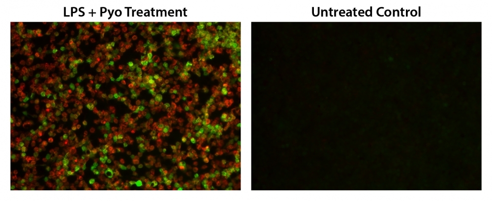
Reactive oxygen species (ROS) are natural byproducts of the normal metabolism of oxygen and play important roles in cell signaling. However, during oxidative stress-related states, ROS levels can increase dramatically. The accumulation of ROS results in significant damage to cell structures. The role of oxidative stress in cardiovascular disease, diabetes, osteoporosis, stroke, inflammatory diseases, a number of neurodegenerative diseases and cancer has been well established. The ROS measurement will help to determine how oxidative stress modulates varied intracellular pathways. Cell Meter™ Fluorimetric ROS Assay Kit uses our unique ROS sensor to quantify ROS in live cells. ROS Green is cell-permeable. It generates the green fluorescence when it reacts with ROS. The kit is an optimized "mix and read" assay format that is compatible with HTS liquid handling instruments. The Cell Meter™ Fluorimetric ROS Assay Kit provides a sensitive, one-step fluorimetric assay to detect intracellular ROS in live cells with one hour incubation. The assay can be performed in a convenient 96-well or 384-well microtiter-plate format and easily adapted to automation without a separation step. Its signal can be easily read using either a fluorescence microplate reader or a fluorescence microscope.

Platform


Fluorescence microscope

ExcitationFITC filter
EmissionFITC filter
Recommended plateBlack wall/clear bottom

Fluorescence microplate reader

Excitation490 nm
Emission525 nm
Cutoff515 nm
Recommended plateBlack wall/clear bottom
Instrument specification(s)Bottom read mode

Components


Example protocol


AT A GLANCE

Protocol summary

  1. Prepare cells in growth medium
  2. Add Amplite™ ROS Green working solution (100 µL/well for a 96- well plate or 25 µL/well for a 384-well plate) 
  3. Stain the cells at 37°C for 60 minutes
  4. Treat the cells with test compounds to induce ROS
  5. Monitor the fluorescence increase (bottom read mode) at Ex/Em= 490/525 nm (Cutoff = 515 nm) or fluorescence microscope with FITC filter set

Important notes
Thaw all the kit components at room temperature before starting the experiment.

PREPARATION OF STOCK SOLUTION

Unless otherwise noted, all unused stock solutions should be divided into single-use aliquots and stored at -20 °C after preparation. Avoid repeated freeze-thaw cycles.

1. Amplite™ ROS Green stock solution (500X):
Add 40 µL of DMSO (Component C) into the vial of Amplite™ ROS Green (Component A) and mix well to make 500X Amplite™ ROS Green stock solution. Protect from light. Note: 20 µL of 500X Amplite™ ROS Green stock solution is enough for 1 plate. For storage, seal tubes tightly.

PREPARATION OF WORKING SOLUTION

Add 20 µL of 500X Amplite™ ROS Green stock solution into 10 mL of Assay Buffer (Component B) and mix well to make Amplite™ ROS Green working solution. Note: This Amplite™ ROS Green working solution is stable for at least 2 hours at room temperature.

For guidelines on cell sample preparation, please visit
https://www.aatbio.com/resources/guides/cell-sample-preparation.html

SAMPLE EXPERIMENTAL PROTOCOL

  1. Add 100 µL/well (96-well plate) or 25 µL/well (384-well plate) of Amplite™ ROS Green working solution into the cell plate.

  2. Incubate the cells in a 5% CO2, 37°C incubator for one hour.

  3. Treat cells with 20 µL of 11X test compounds (96-well plate) or 10 µL of 6X test compounds (384-well plate) in your desired buffer (such as PBS or HHBS). For control wells (untreated cells), add the corresponding amount of compound buffer.

  4. To induce ROS, incubate the cell plate at room temperature or in a 5% CO2, 37°C incubator for at least 15 minutes or a desired period of time (30 minutes for Hela cells treated with 1 mM H2O2).

  5. Monitor the fluorescence increase with a fluorescence microplate reader (bottom read mode) at Ex/Em = 490/525 nm (Cutoff = 515 nm) or observe cells using a fluorescence microscope with FITC filter set.

Images


Citations


View all 33 citations: Citation Explorer
Linoleic acid exhibits anti-proliferative and anti-invasive activities in endometrial cancer cells and a transgenic model of endometrial cancer
Authors: Qiu, Jianqing and Zhao, Ziyi and Suo, Hongyan and Paraghamian, Sarah E and Hawkins, Gabrielle M and Sun, Wenchuan and Zhang, Xin and Hao, Tianran and Deng, Beor and Shen, Xiaochang and others,
Journal: Cancer Biology \& Therapy (2024): 2325130
Reduced expression of phosphorylated ataxia-telangiectasia mutated gene is related to poor prognosis and gemcitabine chemoresistance in pancreatic cancer
Authors: Xun, Jingyu and Ohtsuka, Hideo and Hirose, Katsuya and Douchi, Daisuke and Nakayama, Shun and Ishida, Masaharu and Miura, Takayuki and Ariake, Kyohei and Mizuma, Masamichi and Nakagawa, Kei and others,
Journal: BMC Cancer (2023): 1--13
Shank3 ameliorates neuronal injury after cerebral ischemia/reperfusion via inhibiting oxidative stress and inflammation
Authors: Zhang, Hongchen and Feng, Yuan and Si, Yanfang and Lu, Chuanhao and Wang, Juan and Wang, Shiquan and Li, Liang and Xie, Wenyu and Yue, Zheming and Yong, Jia and others,
Journal: Redox Biology (2023): 102983
Anti-Inflammatory Effects of $\beta$-Cryptoxanthin on 5-Fluorouracil-Induced Cytokine Expression in Human Oral Mucosal Keratinocytes
Authors: Yamanobe, Hironaka and Yamamoto, Kenta and Kishimoto, Saki and Nakai, Kei and Oseko, Fumishige and Yamamoto, Toshiro and Mazda, Osam and Kanamura, Narisato
Journal: Molecules (2023): 2935
Obstructive sleep apnea-increased DEC1 regulates systemic inflammation and oxidative stress that promotes development of pulmonary arterial hypertension
Authors: Li, Xiaoming and Zhang, Xiang and Hou, Xiaozhi and Bing, Xin and Zhu, Fangyuan and Wu, Xinhao and Guo, Na and Zhao, Hui and Xu, Fenglei and Xia, Ming
Journal: Apoptosis (2022): 1--15
ONC206 has anti-tumorigenic effects in human ovarian cancer cells and in a transgenic mouse model of high-grade serous ovarian cancer
Authors: Tucker, Katherine and Yin, Yajie and Staley, Stuart-Allison and Zhao, Ziyi and Fang, Ziwei and Fan, Yali and Zhang, Xin and Suo, Hongyan and Sun, Wenchuan and Prabhu, Varun Vijay and others,
Journal: American Journal of Cancer Research (2022): 521
Reversal of multidrug resistance by Fissistigma latifolium--derived chalconoid 2-hydroxy-4, 5, 6-trimethoxydihydrochalcone in cancer cell lines overexpressing human P-glycoprotein
Authors: Teng, Yu-Ning and Hung, Chin-Chuan and Kao, Pei-Heng and Chang, Ying-Tzu and Lan, Yu-Hsuan
Journal: Biomedicine \& Pharmacotherapy (2022): 113832
GRP75-faciliated Mitochondria-associated ER Membrane (MAM) Integrity controls Cisplatin-resistance in Ovarian Cancer Patients
Authors: Li, Jing and Qi, Fangzheng and Su, Huishan and Zhang, Chuanshan and Zhang, Qing and Chen, Ying and Chen, Ping and Su, Linjia and Chen, Yanan and Yang, Yuqi and others,
Journal: International journal of biological sciences (2022): 2914
The Abnormal Proliferation of Hepatocytes is Associated with MC-LR and C-Terminal Truncated HBX Synergistic Disturbance of the Redox Balance
Authors: Cai, Dong-Mei and Mei, Fan-Biao and Zhang, Chao-Jun and An, San-Chun and Lv, Rui-Bo and Ren, Guan-Hua and Xiao, Chan-Chan and Long, Long and Huang, Tian-Ren and Deng, Wei
Journal: Journal of Hepatocellular Carcinoma (2022): 1229--1246
Antineoplastic Effects and Mechanisms of a New RGD Chimeric Peptide from Bullfrog Skin on the Proliferation and Apoptosis of B16F10 Cells
Authors: Jiang, Xuan and Zhang, Xin and Fu, Chao and Zhao, Ruili and Jin, Tianming and Liu, Mengyue and Pan, Chenhao and Li, Liu An and Ma, Jifei and Yu, Enyuan and others,
Journal: The Protein Journal (2021): 1--12

References


View all 48 references: Citation Explorer
Automatic flow injection based methodologies for determination of scavenging capacity against biologically relevant reactive species of oxygen and nitrogen
Authors: Magalhaes LM, Lucio M, Segundo MA, Reis S, Lima JL.
Journal: Talanta (2009): 1219
Diabetes and the impairment of reproductive function: possible role of mitochondria and reactive oxygen species
Authors: Amaral S, Oliveira PJ, Ramalho-Santos J.
Journal: Curr Diabetes Rev (2008): 46
Virion disruption by ozone-mediated reactive oxygen species
Authors: Murray BK, Ohmine S, Tomer DP, Jensen KJ, Johnson FB, Kirsi JJ, Robison RA, O'Neill KL.
Journal: J Virol Methods (2008): 74
The role of mitochondria in reactive oxygen species metabolism and signaling
Authors: Starkov AA., undefined
Journal: Ann N Y Acad Sci (2008): 37
Sensitive determination of reactive oxygen species by chemiluminescence methods and their application to biological samples and health foods
Authors: Wada M., undefined
Journal: Yakugaku Zasshi (2008): 1031
Reactive oxygen species and yeast apoptosis
Authors: Perrone GG, Tan SX, Dawes IW.
Journal: Biochim Biophys Acta (2008): 1354
Measurement of reactive oxygen species in cells and mitochondria
Authors: Armstrong JS, Whiteman M.
Journal: Methods Cell Biol (2007): 355
Role of reactive oxygen species in mediating hepatic ischemia-reperfusion injury and its therapeutic applications in liver transplantation
Authors: Zhang W, Wang M, Xie HY, Zhou L, Meng XQ, Shi J, Zheng S.
Journal: Transplant Proc (2007): 1332
Superoxide and derived reactive oxygen species in the regulation of hypoxia-inducible factors
Authors: Gorlach A, Kietzmann T.
Journal: Methods Enzymol (2007): 421
Reactive oxygen species and superoxide dismutases: role in joint diseases
Authors: Afonso V, Champy R, Mitrovic D, Collin P, Lomri A.
Journal: Joint Bone Spine (2007): 324