Amplite® Universal Fluorimetric Protease Activity Assay Kit *Green Fluorescence*
Ordering information
Price | |
Catalog Number | |
Unit Size | |
Quantity |
Additional ordering information
Telephone | 1-800-990-8053 |
Fax | 1-800-609-2943 |
sales@aatbio.com | |
International | See distributors |
Bulk request | Inquire |
Custom size | Inquire |
Shipping | Standard overnight for United States, inquire for international |
Spectral properties
Correction Factor (280 nm) | 0.35 |
Extinction coefficient (cm -1 M -1) | 73000 |
Excitation (nm) | 491 |
Emission (nm) | 516 |
Quantum yield | 0.92 |
Storage, safety and handling
H-phrase | H303, H313, H333 |
Hazard symbol | XN |
Intended use | Research Use Only (RUO) |
R-phrase | R20, R21, R22 |
UNSPSC | 12352200 |
Overview | ![]() ![]() |
See also: Amplite® Reagents and Assay Kits, Proteases
Correction Factor (280 nm) 0.35 | Extinction coefficient (cm -1 M -1) 73000 | Excitation (nm) 491 | Emission (nm) 516 | Quantum yield 0.92 |
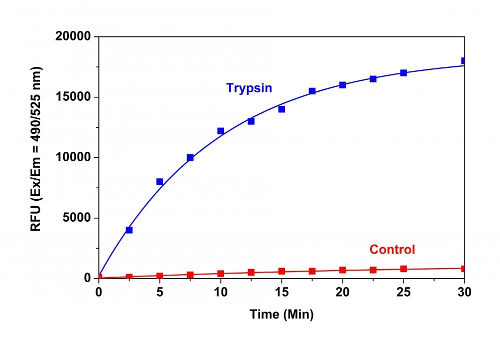
Monitoring of various protease activities has become a routine task for many biological laboratories. Our Amplite® Universal Fluorimetric Protease Activity Assay Kits are an ideal choice for performing routine assays necessary during the isolation of proteases, or for identifying the presence of contaminating proteases in protein samples. The kits use fluorescent casein conjugates that are proven to be a generic substrate for a broad spectrum of proteases. In the intact substrate, casein is heavily labeled with a fluorescent dye, resulting in significant fluorescence quenching. Protease-catalyzed hydrolysis relieves its quenching effect, yielding brightly fluorescent dye-labeled short peptides. The increase in fluorescence intensity is directly proportional to protease activity. The kits provide all the essential components with an optimized "mix & read" protocol that can be easily automated to HTS instruments.
Platform
Fluorescence microplate reader
Excitation | 490 nm |
Emission | 525 nm |
Cutoff | 515 nm |
Recommended plate | Solid black |
Components
Example protocol
AT A GLANCE
Protocol Summary
Measuring protease activity in test samples (Protocol A)- Prepare protease substrate solution (50 µL)
- Add substrate control, positive control or test samples (50 µL)
- Skip incubation for kinetic reading or incubate for 30 to 60 minutes for end point reading
- Monitor fluorescence intensity at Ex/Em = 490/525 nm
Protocol Summary
Screening protease inhibitors using a purified enzyme (Protocol B)- Prepare protease substrate solution (10 µL)
- Add substrate control, positive control, vehicle control or test samples (90 µL)
- Skip incubation for kinetic reading or incubate for 30 to 60 minutes for end point reading
- Monitor fluorescence intensity at Ex/Em = 490/525 nm
PREPARATION OF WORKING SOLUTION
1. Protease substrate solution (For protocol A)
Dilute Protease Substrate (Component A) at 1:100 in 2X assay buffer (Component C). Use 50 µL of protease substrate solution per assay in a 96-well plate. Note The 2X Assay Buffer (Component C) is designed for detecting the activity of chymotrypsin, trypsin, thermolysin, proteinase K, protease XIV, and human leukocyte elastase. For other proteases, please refer to Table 1 below for the appropriate assay buffer formula.
2. Trypsin dilution (For protocol A)
Dilute Trypsin (5 U/µL, Component B) at 1:50 in de-ionized water to get a concentration of 0.1 U/µL.3. Assay Buffer (1X) (For protocol B)
Add 5 mL de-ionized water into 5 mL of 2X Assay Buffer (Component C).4. Protease substrate solution (For protocol B)
Dilute Protease Substrate (Component A) at 1:20 in 1X assay buffer. Use 10 µL/well of protease substrate solution for a 96-well plate. Note The 2X assay buffer (Component C) is designed for detecting the activity of chymotrypsin, trypsin, thermolysin, proteinase K, protease XIV, and human leukocyte elastase. For other proteases, please refer to Table 1 below for the appropriate assay buffer formula.
5. Protease dilution (For protocol B)
Dilute the protease in 1X assay buffer to a concentration of 500 - 1000 nM (For Trypsin 50-100 U/mL). Each well will need 10 µL of protease dilution. Prepare an appropriate amount for all the test samples and extra for the positive control and vehicle control wells.Table 1.Assay buffer formulas for proteases. For protocol A, 2X assay buffer is needed. For protocol B, 1X assay buffer is needed.
Protease | 1X Assay Buffer |
Cathepsin D | 20 mM Sodium Citrate, pH 3.0 |
Papain | 20 mM sodium acetate, 20 mM cysteine, 2 mM EDTA, pH 6.5 |
PAE | 20 mM sodium phosphate, pH 8.0 |
Pepsin | 10 mM HCl, pH 2.0 |
Porcine pancreas elastase | 10 mM Tris-HCl, pH 8.8 |
Subtilisin | 20 mM potassium phosphate buffer, pH 7.6, 150 mM NaCl |
SAMPLE EXPERIMENTAL PROTOCOL
Protocol A: Measure protease activity in test samples
Table 1.Layout of the substrate control, positive control, and test samples in a 96-well microplate. SC=Substrate Control, PC =Positive Control, TS=Test Samples.
SC | SC | ... | ... |
PC | PC | ... | ... |
TS | TS | ||
... | ... | ||
Well | Volume | Reagent |
SC | 50 µL | De-ionized water |
PC | 50 µL | Trypsin dilution |
TS | 50 µL | Protease-containing solution |
- Add 50 µL of protease substrate solution (Protocol A) to all the wells in the assay plate. Mix the reagents well.
- Monitor the fluorescence increase with a fluorescence plate reader at Ex/Em = 490/525 nm. For kinetic reading: Immediately start measuring fluorescence intensity continuously and record data every 5 minutes for 30 minutes. For end-point reading: Incubate the reaction at a desired temperature for 30 to 60 minutes, protected from light. Then measure the fluorescence intensity.
Protocol B: Screening protease inhibitors using a purified enzyme
Table 3.Layout of the samples in a 96-well microplate. SC=Substrate Control, PC= Positive Control, VC=Vehicle Control, TS=Test Samples. It’s recommended to test at least three different concentrations of each test compound. All the test samples should be done in duplicates or triplicates.SC | SC | ... | ... |
PC | PC | ... | ... |
VC | VC | ||
TS | TS | ||
... | ... | ||
Well | Volume | Reagent |
SC | 90 µL | Assay Buffer (1X) (90 µL) |
PC | 90 µL | Assay Buffer (1X) (80 µL) Protease dilution (10 µL) |
VC | 90 µL | Vehicle (X µL) Assay Buffer (80 - X µL) Protease dilution (10 µL) |
TS | 90 µL | Test compound (X µL) Assay Buffer (1X) (80 - X µL) Protease dilution (10 µL) |
- Add 10 µL of protease substrate solution (Protocol B) into the wells of positive control (PC), vehicle control (VC), and test sample (TS). Mix the reagents well.
- Monitor the fluorescence intensity with a fluorescence plate reader at Ex/Em = 490 /525 nm. For kinetic reading: Immediately start measuring fluorescence intensity continuously and record data every 5 minutes for 30 minutes. For end-point reading: Incubate the reaction at a desired temperature for 30 to 60 minutes, protected from light. Then measure the fluorescence intensity.
Spectrum
Open in Advanced Spectrum Viewer


Spectral properties
Correction Factor (280 nm) | 0.35 |
Extinction coefficient (cm -1 M -1) | 73000 |
Excitation (nm) | 491 |
Emission (nm) | 516 |
Quantum yield | 0.92 |
Product Family
Name | Excitation (nm) | Emission (nm) | Extinction coefficient (cm -1 M -1) | Correction Factor (260 nm) | Correction Factor (280 nm) |
Amplite® Universal Fluorimetric Protease Activity Assay Kit *Red Fluorescence* | 552 | 578 | 90000 | 0.32 | 0.178 |
Images

Figure 1. Trypsin protease activity was analyzed by Amplite® Universal Fluorimetric Protease Activity Assay Kit. Protease substrate was incubated with 1 unit trypsin in the kit assay buffer. The control wells had protease substrate only (without trypsin). The fluorescence signal was measured starting from time 0 when trypsin was added. Samples were done in triplicates.

Figure 2. Protease activity of keratinocytes treated with Cry j1 with or without tranexamic acid, SBTI and bivalirudin. (a) Application of Cry j1 (100 ng/ml) to cultured human keratinocytes at time 0 s induced a rapid, transient increase of protease activity (red line). After application of BSS(+) alone (control), the fluorescence level gradually decreased (gray line). The transient increase was blocked by tranexamic acid (blue line), SBTI (yellow line) or bivalirudin (purple line). The vertical scale is normalized by the fluorescence at time 0. (b) Quantitation of fluorescence change within 1 min after application (n = 30–50 cells). A significant difference was observed between the control and Cry j1 application groups. Tranexamic acid, SBTI and bivalirudin each blocked the increase. Similar results were obtained in three independent experiments. Bars and lines represent mean ± SD. (c) Application of Cry j1 (100 ng/ml) to the cells treated with scramble RNA at time 0 s induced a rapid, transient increase of protease activity (gray line), while the application of the same amount of Cry j1 to the thrombin siRNA-treated cells induced a low transient increase (cyan line). The vertical scale is normalized by the fluorescence at time 0. (d) Quantitation of fluorescence change within 1 min after application (n = 30–50 cells). A significant difference was observed between the thrombin siRNA-treated cells and control. Similar results were obtained in three independent experiments. Bars and lines represent mean ± SD. Source: Tranexamic acid blocks the thrombin-mediated delay of epidermal permeability barrier recovery induced by the cedar pollen allergen, Cry j1 by Nakanishi et al., Scientific Reports, Oct. 2018.
Citations
View all 10 citations: Citation Explorer
Comparative assessment of commercially available wound gels in ex vivo human skin reveals major differences in immune response-modulatory effects
Authors: Seiser, S and Cerbu, D and Gallhofer, A and Matiasek, J and Elbe-B{\"u}rger, A
Journal: Scientific reports (2022): 1--9
Authors: Seiser, S and Cerbu, D and Gallhofer, A and Matiasek, J and Elbe-B{\"u}rger, A
Journal: Scientific reports (2022): 1--9
Optimization of Isolation Method for Extracellular Vesicles from Pancreatic Juice and Impact of Protease Activity
Authors: Tsutsumi, Koichiro and Ueta, Eijiro and Kato, Hironari and Matsumoto, Kazuyuki and Horiguchi, Shigeru and Okada, Hiroyuki
Journal: Digestive diseases and sciences (2022): 1--8
Authors: Tsutsumi, Koichiro and Ueta, Eijiro and Kato, Hironari and Matsumoto, Kazuyuki and Horiguchi, Shigeru and Okada, Hiroyuki
Journal: Digestive diseases and sciences (2022): 1--8
A novel polyethylene glycol (PEG)-drug conjugate of Venetoclax, a Bcl-2 inhibitor, for treatment of acute myeloid leukemia (AML)
Authors: Ando, Hidenori and Murakami, Yuta and Eshima, Kiyoshi and Ishida, Tatsuhiro
Journal: Cancer Reports (2021): e1485
Authors: Ando, Hidenori and Murakami, Yuta and Eshima, Kiyoshi and Ishida, Tatsuhiro
Journal: Cancer Reports (2021): e1485
Chondroitin sulfate--mediated N-cadherin/$\beta$-catenin signaling is associated with basal-like breast cancer cell invasion
Authors: Nadanaka, Satomi and Kinouchi, Hiroki and Kitagawa, Hiroshi
Journal: Journal of Biological Chemistry (2018): 444--465
Authors: Nadanaka, Satomi and Kinouchi, Hiroki and Kitagawa, Hiroshi
Journal: Journal of Biological Chemistry (2018): 444--465
IL-33, IL-25 and TSLP contribute to development of fungal-associated protease-induced innate-type airway inflammation
Authors: Hiraishi, Yoshihisa and Yamaguchi, Sachiko and Yoshizaki, Takamichi and Nambu, Aya and Shimura, Eri and Takamori, Ayako and Narushima, Seiko and Nakanishi, Wakako and Asada, Yosuke and Numata, Takafumi and others, undefined
Journal: Scientific reports (2018): 18052
Authors: Hiraishi, Yoshihisa and Yamaguchi, Sachiko and Yoshizaki, Takamichi and Nambu, Aya and Shimura, Eri and Takamori, Ayako and Narushima, Seiko and Nakanishi, Wakako and Asada, Yosuke and Numata, Takafumi and others, undefined
Journal: Scientific reports (2018): 18052
Tranexamic acid blocks the thrombin-mediated delay of epidermal permeability barrier recovery induced by the cedar pollen allergen, Cry j1
Authors: Nakanishi, S and Kumamoto, J and Denda, M
Journal: Scientific reports (2018): 15610
Authors: Nakanishi, S and Kumamoto, J and Denda, M
Journal: Scientific reports (2018): 15610
Eosinophil extracellular trap cell death--derived DNA traps: Their presence in secretions and functional attributes
Authors: Ueki, Shigeharu and Konno, Yasunori and Takeda, Masahide and Moritoki, Yuki and Hirokawa, Makoto and Matsuwaki, Yoshinori and Honda, Kohei and Ohta, Nobuo and Yamamoto, Shiori and Takagi, Yuri and others, undefined
Journal: Journal of Allergy and Clinical Immunology (2016): 258--267
Authors: Ueki, Shigeharu and Konno, Yasunori and Takeda, Masahide and Moritoki, Yuki and Hirokawa, Makoto and Matsuwaki, Yoshinori and Honda, Kohei and Ohta, Nobuo and Yamamoto, Shiori and Takagi, Yuri and others, undefined
Journal: Journal of Allergy and Clinical Immunology (2016): 258--267
Japanese Cedar (Cryptomeria japonica) pollen allergen induces elevation of intracellular calcium in human keratinocytes and impairs epidermal barrier function of human skin ex vivo
Authors: Kumamoto, Junichi and Tsutsumi, Moe and Goto, Makiko and Nagayama, Masaharu and Denda, Mitsuhiro
Journal: Archives of dermatological research (2016): 49--54
Authors: Kumamoto, Junichi and Tsutsumi, Moe and Goto, Makiko and Nagayama, Masaharu and Denda, Mitsuhiro
Journal: Archives of dermatological research (2016): 49--54
Impact of silk biomaterial structure on proteolysis
Authors: Brown, Joseph and Lu, Chia-Li and Coburn, Jeannine and Kaplan, David L
Journal: Acta biomaterialia (2015): 212--221
Authors: Brown, Joseph and Lu, Chia-Li and Coburn, Jeannine and Kaplan, David L
Journal: Acta biomaterialia (2015): 212--221
Incorporation of proteinase inhibitors into silk-based delivery devices for enhanced control of degradation and drug release
Authors: Pritchard, Eleanor M and Valentin, Thomas and Boison, Detlev and Kaplan, David L
Journal: Biomaterials (2011): 909--918
Authors: Pritchard, Eleanor M and Valentin, Thomas and Boison, Detlev and Kaplan, David L
Journal: Biomaterials (2011): 909--918
References
View all 30 references: Citation Explorer
Transient kinetic experiments demonstrate the existence of a unique catalytic enzyme form in the peptide-stimulated ATPase mechanism of Escherichia coli Lon protease
Authors: Vineyard D, Zhang X, Lee I.
Journal: Biochemistry (2006): 11432
Authors: Vineyard D, Zhang X, Lee I.
Journal: Biochemistry (2006): 11432
Highly stable glycosylated serine protease from the medicinal plant Euphorbia milii
Authors: Yadav SC, P and e M, Jagannadham MV.
Journal: Phytochemistry (2006): 1414
Authors: Yadav SC, P and e M, Jagannadham MV.
Journal: Phytochemistry (2006): 1414
Effects of Pseudomonas fluorescens M3/6 bacterial protease on plasmin system and plasminogen activation
Authors: Frohbieter KA, Ismail B, Nielsen SS, Hayes KD.
Journal: J Dairy Sci (2005): 3392
Authors: Frohbieter KA, Ismail B, Nielsen SS, Hayes KD.
Journal: J Dairy Sci (2005): 3392
Fibrillar amyloid beta-protein inhibits the activity of high molecular weight brain protease and trypsin
Authors: Chauhan V, Sheikh AM, Chauhan A, Spivack WD, Fenko MD, Malik MN.
Journal: J Alzheimers Dis (2005): 37
Authors: Chauhan V, Sheikh AM, Chauhan A, Spivack WD, Fenko MD, Malik MN.
Journal: J Alzheimers Dis (2005): 37
Characterization of a novel and specific inhibitor for the pro-apoptotic protease Omi/HtrA2
Authors: Cilenti L, Lee Y, Hess S, Srinivasula S, Park KM, Junqueira D, Davis H, Bonventre JV, Alnemri ES, Zervos AS.
Journal: J Biol Chem (2003): 11489
Authors: Cilenti L, Lee Y, Hess S, Srinivasula S, Park KM, Junqueira D, Davis H, Bonventre JV, Alnemri ES, Zervos AS.
Journal: J Biol Chem (2003): 11489
Measurement of protease activity of live Uronema marinun (Ciliata: Scuticociliatida) by fluorescence polarization
Authors: Lee EH, Kim CS, Cho JB, Ahn KJ, Kim KH.
Journal: Dis Aquat Organ (2003): 85
Authors: Lee EH, Kim CS, Cho JB, Ahn KJ, Kim KH.
Journal: Dis Aquat Organ (2003): 85
Mg2+-linked oligomerization modulates the catalytic activity of the Lon (La) protease from Mycobacterium smegmatis
Authors: Rudyak SG, Brenowitz M, Shrader TE.
Journal: Biochemistry (2001): 9317
Authors: Rudyak SG, Brenowitz M, Shrader TE.
Journal: Biochemistry (2001): 9317
Directed polar secretion of protease from single cells of Vibrio cholerae via the type II secretion pathway
Authors: Scott ME, Dossani ZY, S and kvist M., undefined
Journal: Proc Natl Acad Sci U S A (2001): 13978
Authors: Scott ME, Dossani ZY, S and kvist M., undefined
Journal: Proc Natl Acad Sci U S A (2001): 13978
Effect of psychrotrophic bacteria and of an isolated protease from Pseudomonas fluorescens M3/6 on the plasmin system of fresh milk
Authors: Fajardo-Lira C, Oria M, Hayes KD, Nielsen SS.
Journal: J Dairy Sci (2000): 2190
Authors: Fajardo-Lira C, Oria M, Hayes KD, Nielsen SS.
Journal: J Dairy Sci (2000): 2190
Expression, purification, and functional analysis of the human serine protease HtrA2
Authors: Savopoulos JW, Carter PS, Turconi S, Pettman GR, Karran EH, Gray CW, Ward RV, Jenkins O, Creasy CL.
Journal: Protein Expr Purif (2000): 227
Authors: Savopoulos JW, Carter PS, Turconi S, Pettman GR, Karran EH, Gray CW, Ward RV, Jenkins O, Creasy CL.
Journal: Protein Expr Purif (2000): 227
Application notes
A Library of Well-Defined and Water-Soluble Poly(alkyl phosphonate)s with Adjustable Hydrolysis
Acetylcholinesterase Inhibitory Activity of Pigment Echinochrome A
Ameliorative Effect of Novel Vitamin Formula with Herbal Extracts on Scopolamine-Induced Alzheimer's Disease
An Increase in Plasma Homovanillic Acid with Cocoa Extract Consumption Is Associated with the Alleviation of Depressive Symptoms in Overweight or Obese Adults
Attenuation of lysyl oxidase and collagen gene expression in keratoconus patient corneal epithelium corresponds to disease severity