logo
AAT Bioquest

Calbryte™ 590 AM

The intracellular calcium flux assay is a widely used method in monitoring signal transduction pathways and high throughput screening of G protein"coupled receptors (GPCRs) and calcium channel targets. Followed by Rhod-2 being introduced in 1989, Rhod-4 and Cal-590 were later developed with improved signal/background ratio, and they became the widely used red fluorescent Ca2+ indicators for confocal microscopy, flow cytometry and high throughput screening applications. In CHO and HEK cells Rhod-4 and Cal-590 have cellular calcium response that are 10 times more sensitive than Rhod-2 AM. However, Cal-590 and Rhod-4 are still less sensitive to calcium in cells than the corresponding green fluorescent calcium indicators (e.g., Fluo-8 and Cal-520). Calbryte™ 590 is a new generation of red fluorescent indicators for the measurement of intracellular calcium. Its greatly improved signal/background ratio and intracellular retention properties make Calbryte™ 590 AM the most robust red fluorescent indicator for evaluating GPCR and calcium channel targets as well as for screening their agonists and antagonists in live cells. Like other dye AM cell loading, Calbryte™ 590 AM ester is non-fluorescent and once gets inside the cell, it is hydrolyzed by intracellular esterase and gets activated. The activated indicator is a polar molecule that is no longer capable of freely diffusing through cell membrane, essentially trapped inside cells.
Example protocol

PREPARATION OF STOCK SOLUTIONS

Unless otherwise noted, all unused stock solutions should be divided into single-use aliquots and stored at -20 °C after preparation. Avoid repeated freeze-thaw cycles

Calbryte™ 590 AM Stock Solution
  1. Prepare a 2 to 5 mM stock solution of Calbryte™ 590 AM in anhydrous DMSO.

    Note: When reconstituted in DMSO, Calbryte™ 590 AM is a clear, colorless solution.

PREPARATION OF WORKING SOLUTION

Calbryte™ 590 AM Working Solution
  1. On the day of the experiment, either dissolve Calbryte™ 590 AM in DMSO or thaw an aliquot of the indicator stock solution to room temperature.

  2. Prepare a 2 to 20 µM Calbryte™ 590 AM working solution in a buffer of your choice (e.g., Hanks and Hepes buffer) with 0.04% Pluronic® F-127. For most cell lines, Calbryte™ 590 AM at a final concentration of 4-5 μM is recommended. The exact concentration of indicators required for cell loading must be determined empirically.

    Note: The nonionic detergent Pluronic® F-127 is sometimes used to increase the aqueous solubility of Calbryte™ 590 AM. A variety of Pluronic® F-127 solutions can be purchased from AAT Bioquest.

    Note: If your cells contain organic anion-transporters, probenecid (1-2 mM) may be added to the dye working solution (final in well concentration will be 0.5-1 mM) to reduce leakage of the de-esterified indicators. A variety of ReadiUse™ Probenecid products, including water-soluble, sodium salt, and stabilized solutions, can be purchased from AAT Bioquest.

SAMPLE EXPERIMENTAL PROTOCOL

Following is our recommended protocol for loading AM esters into live cells. This protocol only provides a guideline and should be modified according to your specific needs.

  1. Prepare cells in growth medium overnight.
  2. On the next day, add 1X Calbryte™ 590 AM working solution to your cell plate.

    Note: If your compound(s) interfere with the serum, replace the growth medium with fresh HHBS buffer before dye-loading.

  3. Incubate the dye-loaded plate in a cell incubator at 37 °C for 30 to 60 minutes.

    Note: Incubating the dye for longer than 1 hour can improve signal intensities in certain cell lines.

  4. Replace the dye working solution with HHBS or buffer of your choice (containing an anion transporter inhibitor, such as 1 mM probenecid, if applicable) to remove any excess probes.
  5. Add the stimulant as desired and simultaneously measure fluorescence using either a fluorescence microscope equipped with a TRITC/Cy3 filter set or a fluorescence plate reader containing a programmable liquid handling system such as an FDSS, FLIPR, or FlexStation, at Ex/Em = 540/590 nm cutoff 570 nm.

Calculators

Common stock solution preparation

Table 1. Volume of DMSO needed to reconstitute specific mass of Calbryte™ 590 AM to given concentration. Note that volume is only for preparing stock solution. Refer to sample experimental protocol for appropriate experimental/physiological buffers.

0.1 mg0.5 mg1 mg5 mg10 mg
1 mM82.05 µL410.25 µL820.499 µL4.102 mL8.205 mL
5 mM16.41 µL82.05 µL164.1 µL820.499 µL1.641 mL
10 mM8.205 µL41.025 µL82.05 µL410.25 µL820.499 µL

Molarity calculator

Enter any two values (mass, volume, concentration) to calculate the third.

Mass (Calculate)Molecular weightVolume (Calculate)Concentration (Calculate)Moles
/=x=

Spectrum

Product family

NameExcitation (nm)Emission (nm)Quantum yield
Calbryte™ 520 AM4935150.751
Calbryte™ 630 AM607624-
Calbryte™-520L AM4935150.751
Calbryte™-520XL AM4935150.751
Cal-590™ AM5745880.621

Citations

View all 54 citations: Citation Explorer
Dual regulation of IP3 receptors by IP3 and PIP2 controls the transition from local to global Ca2+ signals
Authors: Ivanova, Adelina and Atakpa-Adaji, Peace and Rao, Shanlin and Marti-Solano, Maria and Taylor, Colin W
Journal: Molecular Cell (2024)
Scar matrix drives Piezo1 mediated stromal inflammation leading to placenta accreta spectrum
Authors: Wenqiang, Du and Novin, Ashkan and Liu, Yamin and Afzal, Junaid and Suhail, Yasir and Liu, Shaofei and Gavin, Nicole R and Jorgensen, Jennifer R and Morosky, Christopher M and Figueroa, Reinaldo and others,
Journal: Nature Communications (2024): 8379
Light Responsive Molecular Probes for In Vivo
Authors: 橋本龍,
Journal: (2024)
AAVS1-targeted, stable expression of ChR2 in human brain organoids for consistent optogenetic control
Authors: Hong, Soojung and Lee, Juhee and Kim, Yunhee and Kim, Eunjee and Shin, Kunyoo
Journal: Bioengineering \& Translational Medicine (2024): e10690

References

View all 53 references: Citation Explorer
A flow cytometric comparison of Indo-1 to fluo-3 and Fura Red excited with low power lasers for detecting Ca(2+) flux
Authors: Bailey S, Macardle PJ.
Journal: J Immunol Methods (2006): 220
Functional fluo-3/AM assay on P-glycoprotein transport activity in L1210/VCR cells by confocal microscopy
Authors: Orlicky J, Sulova Z, Dovinova I, Fiala R, Zahradnikova A, Jr., Breier A.
Journal: Gen Physiol Biophys (2004): 357
Comparison of human recombinant adenosine A2B receptor function assessed by Fluo-3-AM fluorometry and microphysiometry
Authors: Patel H, Porter RH, Palmer AM, Croucher MJ.
Journal: Br J Pharmacol (2003): 671
Measurement of the dissociation constant of Fluo-3 for Ca2+ in isolated rabbit cardiomyocytes using Ca2+ wave characteristics
Authors: Loughrey CM, MacEachern KE, Cooper J, Smith GL.
Journal: Cell Calcium (2003): 1
A sensitive method for the detection of foot and mouth disease virus by in situ hybridisation using biotin-labelled oligodeoxynucleotides and tyramide signal amplification
Authors: Zhang Z, Kitching P.
Journal: J Virol Methods (2000): 187
Page updated on October 12, 2024

Ordering information

Price
AvailabilityIn stock
Unit size
2x50 ug
10x50 ug
1 mg
Catalog Number
Quantity
Add to cart

Additional ordering information

Telephone1-800-990-8053
Fax1-800-609-2943
Emailsales@aatbio.com
InternationalSee distributors
Bulk requestInquire
Custom sizeInquire
Technical SupportContact us
Purchase orderSend to sales@aatbio.com
ShippingStandard overnight for United States, inquire for international
Request quotation

Physical properties

Dissociation constant (Kd, nM)1400

Molecular weight

1218.77

Solvent

DMSO

Spectral properties

Excitation (nm)

581

Emission (nm)

593

Storage, safety and handling

Certificate of OriginDownload PDF
H-phraseH303, H313, H333
Hazard symbolXN
Intended useResearch Use Only (RUO)
R-phraseR20, R21, R22

Storage

Freeze (< -15 °C); Minimize light exposure
UNSPSC12352200

Platform

Fluorescence microscope

ExcitationTRITC, Cy3
EmissionTRITC, Cy3
Recommended plateBlack wall, clear bottom

Fluorescence microplate reader

Excitation540
Emission590
Cutoff570
Recommended plateBlack wall, clear bottom
Instrument specification(s)Bottom read mode, Programmable liquid handling
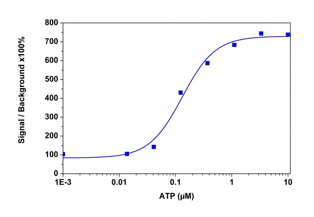
An ATP&nbsp;dose-response was measured in CHO-K1 cells with Calbryte&trade; 590 AM. CHO-K1 cells were seeded overnight at 50,000 cells/100 &micro;L/well in a 96-well black wall/clear bottom costar plate. 100 &micro;L of 10 &micro;g/ml Calbryte&trade; 590 AM in HH Buffer with probenecid was added and incubated for 60 min at 37&deg;C. Dye loading solution was then removed and replaced with 200 &micro;L HH Buffer/well. ATP&nbsp;(50 &micro;L/well) was added by FlexStation 3 to achieve the final indicated concentrations.