Calbryte™ 590 AM
Overview | ![]() ![]() |
Molecular weight 1218.77 | Dissociation constant (Kd, nM) 1400 | Excitation (nm) 581 | Emission (nm) 593 |
The intracellular calcium flux assay is a widely used method in monitoring signal transduction pathways and high throughput screening of G protein"coupled receptors (GPCRs) and calcium channel targets. Followed by Rhod-2 being introduced in 1989, Rhod-4 and Cal-590 were later developed with improved signal/background ratio, and they became the widely used red fluorescent Ca2+ indicators for confocal microscopy, flow cytometry and high throughput screening applications. In CHO and HEK cells Rhod-4 and Cal-590 have cellular calcium response that are 10 times more sensitive than Rhod-2 AM. However, Cal-590 and Rhod-4 are still less sensitive to calcium in cells than the corresponding green fluorescent calcium indicators (e.g., Fluo-8 and Cal-520). Calbryte™ 590 is a new generation of red fluorescent indicators for the measurement of intracellular calcium. Its greatly improved signal/background ratio and intracellular retention properties make Calbryte™ 590 AM the most robust red fluorescent indicator for evaluating GPCR and calcium channel targets as well as for screening their agonists and antagonists in live cells. Like other dye AM cell loading, Calbryte™ 590 AM ester is non-fluorescent and once gets inside the cell, it is hydrolyzed by intracellular esterase and gets activated. The activated indicator is a polar molecule that is no longer capable of freely diffusing through cell membrane, essentially trapped inside cells.
Platform
Fluorescence microscope
Excitation | TRITC/Cy3 |
Emission | TRITC/Cy3 |
Recommended plate | Black wall/clear bottom |
Fluorescence microplate reader
Excitation | 540 |
Emission | 590 |
Cutoff | 570 |
Recommended plate | Black wall/clear bottom |
Instrument specification(s) | Bottom read mode/Programmable liquid handling |
Example protocol
PREPARATION OF STOCK SOLUTIONS
Unless otherwise noted, all unused stock solutions should be divided into single-use aliquots and stored at -20 °C after preparation. Avoid repeated freeze-thaw cycles.
Calbryte™ 590 AM Stock Solution
Prepare a 2 to 5 mM stock solution of Calbryte™ 590 AM in high-quality, anhydrous DMSO.PREPARATION OF WORKING SOLUTION
Calbryte™ 590 AM Working Solution
On the day of the experiment, either dissolve Calbryte™ 590 AM in DMSO or thaw an aliquot of the indicator stock solution to room temperature. Prepare a dye working solution of 2 to 20 µM in a buffer of your choice (e.g., Hanks and Hepes buffer) with 0.04% Pluronic® F-127. For most cell lines, Calbryte™ 590 AM at a final concentration of 4-5 μM is recommended. The exact concentration of indicators required for cell loading must be determined empirically.Note The nonionic detergent Pluronic® F-127 is sometimes used to increase the aqueous solubility of Calbryte™ 590 AM. A variety of Pluronic® F-127 solutions can be purchased from AAT Bioquest.
Note If your cells contain organic anion-transporters, probenecid (1-2 mM) may be added to the dye working solution (final in well concentration will be 0.5-1 mM) to reduce leakage of the de-esterified indicators. A variety of ReadiUse™ probenecid products, including water-soluble, sodium salt, and stabilized solution, can be purchased from AAT Bioquest.
SAMPLE EXPERIMENTAL PROTOCOL
Following is our recommended protocol for loading AM esters into live cells. This protocol only provides a guideline and should be modified according to your specific needs.
- Prepare cells in growth medium overnight.
- On the next day, add 1X Calbryte™ 590 AM working solution into your cell plate.
Note If your compound(s) interfere with the serum, replace the growth medium with fresh HHBS buffer before dye-loading. - Incubate the dye-loaded plate in a cell incubator at 37 °C for 30 to 60 minutes.
Note Incubating the dye for longer than 1 hour can improve signal intensities in certain cell lines. - Replace the dye working solution with HHBS or buffer of your choice (containing an anion transporter inhibitor, such as 1 mM probenecid, if applicable) to remove any excess probes.
- Add the stimulant as desired and simultaneously measure fluorescence using either a fluorescence microscope equipped with a TRITC/Cy3 filter set or a fluorescence plate reader containing a programmable liquid handling system such as an FDSS, FLIPR, or FlexStation, at Ex/Em = 540/590 nm cutoff 570 nm.
Calculators
Common stock solution preparation
Table 1. Volume of DMSO needed to reconstitute specific mass of Calbryte™ 590 AM to given concentration. Note that volume is only for preparing stock solution. Refer to sample experimental protocol for appropriate experimental/physiological buffers.
0.1 mg | 0.5 mg | 1 mg | 5 mg | 10 mg | |
1 mM | 82.05 µL | 410.25 µL | 820.499 µL | 4.102 mL | 8.205 mL |
5 mM | 16.41 µL | 82.05 µL | 164.1 µL | 820.499 µL | 1.641 mL |
10 mM | 8.205 µL | 41.025 µL | 82.05 µL | 410.25 µL | 820.499 µL |
Molarity calculator
Enter any two values (mass, volume, concentration) to calculate the third.
Mass (Calculate) | Molecular weight | Volume (Calculate) | Concentration (Calculate) | Moles | ||||
/ | = | x | = |
Product family
Name | Excitation (nm) | Emission (nm) | Quantum yield |
Calbryte™ 520 AM | 493 | 515 | 0.751 |
Calbryte™ 630 AM | 607 | 624 | - |
Calbryte™-520L AM | 493 | 515 | 0.751 |
Calbryte™-520XL AM | 493 | 515 | 0.751 |
Cal-590™ AM | 574 | 588 | 0.621 |
Images
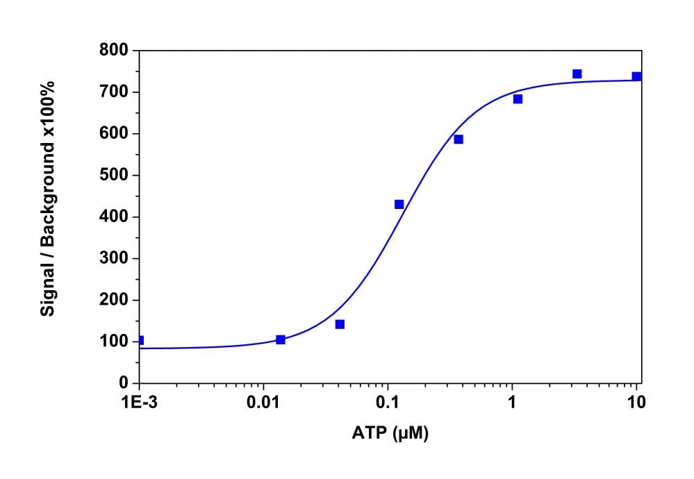
Figure 1. An ATP dose-response was measured in CHO-K1 cells with Calbryte™ 590 AM. CHO-K1 cells were seeded overnight at 50,000 cells/100 µL/well in a 96-well black wall/clear bottom costar plate. 100 µL of 10 µg/ml Calbryte™ 590 AM in HH Buffer with probenecid was added and incubated for 60 min at 37°C. Dye loading solution was then removed and replaced with 200 µL HH Buffer/well. ATP (50 µL/well) was added by FlexStation 3 to achieve the final indicated concentrations.
Citations
View all 32 citations: Citation Explorer
Dissociation of inositol 1, 4, 5-trisphosphate from IP3 receptors contributes to termination of Ca2+ puffs
Authors: Smith, Holly A and Taylor, Colin W
Journal: Journal of Biological Chemistry (2023): 102871
Authors: Smith, Holly A and Taylor, Colin W
Journal: Journal of Biological Chemistry (2023): 102871
The influence of spontaneous and visual activity on the development of direction selectivity maps in mouse retina
Authors: Tiriac, Alexandre and Bistrong, Karina and Pitcher, Miah N and Tworig, Joshua M and Feller, Marla B
Journal: Cell reports (2022): 110225
Authors: Tiriac, Alexandre and Bistrong, Karina and Pitcher, Miah N and Tworig, Joshua M and Feller, Marla B
Journal: Cell reports (2022): 110225
Microglia modulate blood flow, neurovascular coupling, and hypoperfusion via purinergic actions
Authors: Cs{\'a}sz{\'a}r, Eszter and L{\'e}n{\'a}rt, Nikolett and Cser{\'e}p, Csaba and K{\"o}rnyei, Zsuzsanna and Fekete, Rebeka and P{\'o}sfai, Bal{\'a}zs and Bal{\'a}zsfi, Di{\'a}na and Hangya, Bal{\'a}zs and Schwarcz, Anett D and Szabadits, Eszter and others,
Journal: Journal of Experimental Medicine (2022)
Neurodegeneration in human brain organoids infected with herpes simplex virus type 1
Authors: Rybak-Wolf, Agnieszka and Wyler, Emanuel and Legnini, Ivano and Loewa, Anna and Gla{\v{z}}ar, Petar and Kim, Seung Joon and Pentimalli, Tancredi Massimo and Martinez, Anna Oliveras and Beyersdorf, Benjamin and Woehler, Andrew and others,
Journal: bioRxiv (2021)
Authors: Rybak-Wolf, Agnieszka and Wyler, Emanuel and Legnini, Ivano and Loewa, Anna and Gla{\v{z}}ar, Petar and Kim, Seung Joon and Pentimalli, Tancredi Massimo and Martinez, Anna Oliveras and Beyersdorf, Benjamin and Woehler, Andrew and others,
Journal: bioRxiv (2021)
Investigating the Molecular Mechanisms of TMEM16F--a Ca2+ Activated Phospholipid Scramblase and Ion Channel
Authors: Le, Trieu Phuong Hai
Journal: (2021)
Authors: Le, Trieu Phuong Hai
Journal: (2021)
KRAP tethers IP3 receptors to actin and licenses them to evoke cytosolic Ca2+ signals
Authors: Thillaiappan, Nagendra Babu and Smith, Holly A and Atakpa-Adaji, Peace and Taylor, Colin W
Journal: Nature communications (2021): 1--13
Authors: Thillaiappan, Nagendra Babu and Smith, Holly A and Atakpa-Adaji, Peace and Taylor, Colin W
Journal: Nature communications (2021): 1--13
Evidence that polyphenols do not inhibit the phospholipid scramblase TMEM16F
Authors: Le, Trieu and Le, Son C and Zhang, Yang and Liang, Pengfei and Yang, Huanghe
Journal: Journal of Biological Chemistry (2020): 12537--12544
Authors: Le, Trieu and Le, Son C and Zhang, Yang and Liang, Pengfei and Yang, Huanghe
Journal: Journal of Biological Chemistry (2020): 12537--12544
A fluorescent sensor for spatiotemporally resolved endocannabinoid dynamics in vitro and in vivo
Authors: Dong, Ao and He, Kaikai and Dudok, Barna and Farrell, Jordan S and Guan, Wuqiang and Liput, Daniel J and Puhl, Henry L and Cai, Ruyi and Duan, Jiali and Albarran, Eddy and others,
Journal: BioRxiv (2020)
Authors: Dong, Ao and He, Kaikai and Dudok, Barna and Farrell, Jordan S and Guan, Wuqiang and Liput, Daniel J and Puhl, Henry L and Cai, Ruyi and Duan, Jiali and Albarran, Eddy and others,
Journal: BioRxiv (2020)
Ca2+ Release by IP3 Receptors Is Required to Orient the Mitotic Spindle
Authors: Lagos-Cabr{\'e}, Raul and Ivanova, Adelina and Taylor, Colin W
Journal: Cell reports (2020): 108483
Authors: Lagos-Cabr{\'e}, Raul and Ivanova, Adelina and Taylor, Colin W
Journal: Cell reports (2020): 108483
TMEM16F phospholipid scramblase mediates trophoblast fusion and placental development
Authors: Zhang, Yang and Le, Trieu and Grabau, Ryan and Mohseni, Zahra and Kim, Hoejeong and Natale, David R and Feng, Liping and Pan, Hua and Yang, Huanghe
Journal: Science advances (2020): eaba0310
Authors: Zhang, Yang and Le, Trieu and Grabau, Ryan and Mohseni, Zahra and Kim, Hoejeong and Natale, David R and Feng, Liping and Pan, Hua and Yang, Huanghe
Journal: Science advances (2020): eaba0310
References
View all 53 references: Citation Explorer
A flow cytometric comparison of Indo-1 to fluo-3 and Fura Red excited with low power lasers for detecting Ca(2+) flux
Authors: Bailey S, Macardle PJ.
Journal: J Immunol Methods (2006): 220
Authors: Bailey S, Macardle PJ.
Journal: J Immunol Methods (2006): 220
Functional fluo-3/AM assay on P-glycoprotein transport activity in L1210/VCR cells by confocal microscopy
Authors: Orlicky J, Sulova Z, Dovinova I, Fiala R, Zahradnikova A, Jr., Breier A.
Journal: Gen Physiol Biophys (2004): 357
Authors: Orlicky J, Sulova Z, Dovinova I, Fiala R, Zahradnikova A, Jr., Breier A.
Journal: Gen Physiol Biophys (2004): 357
Comparison of human recombinant adenosine A2B receptor function assessed by Fluo-3-AM fluorometry and microphysiometry
Authors: Patel H, Porter RH, Palmer AM, Croucher MJ.
Journal: Br J Pharmacol (2003): 671
Authors: Patel H, Porter RH, Palmer AM, Croucher MJ.
Journal: Br J Pharmacol (2003): 671
Measurement of the dissociation constant of Fluo-3 for Ca2+ in isolated rabbit cardiomyocytes using Ca2+ wave characteristics
Authors: Loughrey CM, MacEachern KE, Cooper J, Smith GL.
Journal: Cell Calcium (2003): 1
Authors: Loughrey CM, MacEachern KE, Cooper J, Smith GL.
Journal: Cell Calcium (2003): 1
A sensitive method for the detection of foot and mouth disease virus by in situ hybridisation using biotin-labelled oligodeoxynucleotides and tyramide signal amplification
Authors: Zhang Z, Kitching P.
Journal: J Virol Methods (2000): 187
Authors: Zhang Z, Kitching P.
Journal: J Virol Methods (2000): 187
Kinetics of onset of mouse sperm acrosome reaction induced by solubilized zona pellucida: fluorimetric determination of loss of pH gradient between acrosomal lumen and medium monitored by dapoxyl (2-aminoethyl) sulfonamide and of intracellular Ca(2+) chang
Authors: Rockwell PL, Storey BT.
Journal: Mol Reprod Dev (2000): 335
Authors: Rockwell PL, Storey BT.
Journal: Mol Reprod Dev (2000): 335
MRP2, a human conjugate export pump, is present and transports fluo 3 into apical vacuoles of Hep G2 cells
Authors: Cantz T, Nies AT, Brom M, Hofmann AF, Keppler D.
Journal: Am J Physiol Gastrointest Liver Physiol (2000): G522
Authors: Cantz T, Nies AT, Brom M, Hofmann AF, Keppler D.
Journal: Am J Physiol Gastrointest Liver Physiol (2000): G522
Use of co-loaded Fluo-3 and Fura Red fluorescent indicators for studying the cytosolic Ca(2+)concentrations distribution in living plant tissue
Authors: Walczysko P, Wagner E, Albrechtova JT.
Journal: Cell Calcium (2000): 23
Authors: Walczysko P, Wagner E, Albrechtova JT.
Journal: Cell Calcium (2000): 23
[Ca2+]i following extrasystoles in guinea-pig trabeculae microinjected with fluo-3 - a comparison with frog skeletal muscle fibres
Authors: Wohlfart B., undefined
Journal: Acta Physiol Scand (2000): 1
Authors: Wohlfart B., undefined
Journal: Acta Physiol Scand (2000): 1
Determination of the intracellular dissociation constant, K(D), of the fluo-3. Ca(2+) complex in mouse sperm for use in estimating intracellular Ca(2+) concentrations
Authors: Rockwell PL, Storey BT.
Journal: Mol Reprod Dev (1999): 418
Authors: Rockwell PL, Storey BT.
Journal: Mol Reprod Dev (1999): 418
Application notes
A Comparison of Fluorescent Red Calcium Indicators for Detecting Intracellular Calcium Mobilization in CHO Cells
Calbryte™ 520, Calbryte™ 590 and Calbryte™ 630 Calcium Detection Reagents
Introducing Calbryte™ Series
A Comparison of Fluorescent Red Calcium Indicators for Detecting Intracellular Calcium Mobilization in CHO Cells
Calbryte™ 520, Calbryte™ 590 and Calbryte™ 630 Calcium Detection Reagents
FAQ
Are there any calcium indicators that don't require probenecid (PBC)?
Are there upgraded trypan blue derivatives for cell viability testing?