logo
AAT Bioquest

Gelite™ Safe DNA Gel Stain *10,000X Water Solution*

<strong>Comparison of DNA detection in 1% agarose gel in TBE buffer using Gelite&trade; Safe, EtBr, and SYBR&reg; Safe.</strong>&nbsp;Two-fold serial dilutions of 1 kb DNA ladder were loaded in amounts of 100 ng, 50 ng, and 25 ng from left to right. Gels were stained for 60 minutes with Gelite&trade; Safe, EtBr, and SYBR&reg; Safe according to the manufacturer's recommended concentrations and imaged using the ChemiDoc&trade; Imaging System (Bio-Rad&reg;). Gels were illuminated using a 300 nm transilluminator fitted with a GelGreen filter.
Comparison of Gelite™ Safe (1:25,000X dilution) and GelRed® (1:10,000X dilution) in precast gel staining using 1% agarose gel in TBE buffer. Two-fold serial dilutions of 1 kb DNA ladder were loaded in the amounts of 100 ng, 50 ng, 25 ng, 12 ng, 6 ng, 3 ng, 1.5 ng, and 0.7 ng from left to right. Gels were imaged using a 300 nm transilluminator in ChemiDoc™ Imaging System (Bio-Rad®).
Stability test on Gelite™ Safe DNA Gel Stain, which was dissolved in water and stored at four different temperatures (-20°C, 4°C, room temperature, and 37°C) for a period of one month. After storage, DNA ladders (100, 50, and 25 ng) were loaded in 1% agarose gel for 60 minutes at 75 V. Gels were incubated with Gelite™ Safe DNA Gel Stain for 1 hour. The image was captured in SYBR green filter set (Exposure time= 1.5 sec).
Stability test on Gelite™ Safe DNA Gel Stain, which was dissolved in water and stored at four different temperatures (-20°C, 4°C, room temperature, and 37°C) for a period of three months. After storage, DNA ladders (100, 50, and 25 ng) were loaded in 1% agarose gel for 60 minutes at 75 V. Gels were incubated with Gelite™ Safe DNA Gel Stain for 1 hour. The image was captured in SYBR green filter set (Exposure time= 1.5 sec).
Comparison of DNA detection in 1% agarose gel in TBE buffer using Gelite™ Safe and GelRed®. Two-fold serial dilutions of 1 kb DNA ladder were loaded in amounts of 100 ng, 50 ng, and 25 ng from left to right. Gels were stained for 60 minutes with Gelite™ Safe and GelRed™ (Biotium) according to the manufacturer's recommended concentrations and imaged using the ChemiDoc™ Imaging System (Bio-Rad®). Gels were illuminated using a 300 nm transilluminator fitted with an EtBr filter set.
Ordering information
Price
Catalog Number
Unit Size
Quantity
Add to cart
Additional ordering information
Telephone1-800-990-8053
Fax1-800-609-2943
Emailsales@aatbio.com
InternationalSee distributors
Bulk requestInquire
Custom sizeInquire
ShippingStandard overnight for United States, inquire for international
Request quotation
Spectral properties
Absorbance (nm)509
Excitation (nm)513
Emission (nm)552
Storage, safety and handling
H-phraseH303, H313, H333
Hazard symbolXN
Intended useResearch Use Only (RUO)
R-phraseR20, R21, R22
UNSPSC12171501

OverviewpdfSDSpdfProtocol


Absorbance (nm)
509
Excitation (nm)
513
Emission (nm)
552
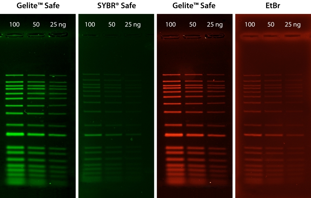
AAT Bioquest is committed to designing our products to be environment-friendly. It is part of how we enable our customers to make the world healthier, cleaner, and safer. Ethidium bromide (EtBr) has been commonly used as a DNA stain for many years. However, EtBr is harmful if swallowed and is very toxic if inhaled. EtBr has been shown to be mutagenic in various tests and is an aquatic toxin. SYBR® Safe was introduced as a safer alternative to EtBr and SYBR® Green, but unfortunately, it is much less sensitive than SYBR® Green. It only has sensitivity comparable to EtBr. Gelite™ Safe has been developed specifically to be less hazardous than EtBr for staining DNA in agarose and acrylamide gels with much higher sensitivity. Gelite™ Safe has greatly improved safety and uncompromised sensitivity. The exceptional sensitivity and strong DNA binding affinity of Gelite™ Safe allows DNA to be stained prior to or post electrophoresis without destaining. In addition to its superior binding properties, Gelite™ Safe is essentially non-fluorescent in the absence of nucleic acids showing very low background fluorescence. Upon binding to nucleic acids, Gelite™ Safe exhibits a considerable fluorescence enhancement by several orders of magnitude greater than that of EtBr. Gelite™ Safe was optimized to be compatible with various instruments, including UV and blue-light transiluminators, gel documentation systems, and laser scanners. It is the first single formulation that can be used in either the green or red channel at your preference. Unlike the membrane-permeant SYBR® Green, which is highly toxic to cells and the environment, the membrane-impermeant properties of Gelite™ Safe make it a much safer and noncytotoxic alternative. Furthermore, Ames testing has confirmed Gelite™ Safe to be significantly less mutagenic than EtBr and SYBR® Green, even at concentrations well above the working concentration used for gel staining. Ames mutagenicity test was performed in a dose-dependent manner for all test dyes pretreated with an S9 fraction from rat liver (SYBR® is a trademark of ThermoFisher).

Platform


Gel Imager

ExcitationUV Transilluminator/Blue laser
EmissionSYBR® filter, GelStar® filter, GelGreen® filter, or GelRed® filter

Example protocol


PREPARATION OF WORKING SOLUTION

Gelite™ Safe working solution
  1. Make 1X Gelite™ Safe working solution by diluting the 10,000X stock reagent with a buffer of your choice in a pH range of 7.5-8.5 (e.g., TAE, TBE, or TE, preferably pH 8.2).

    Note: Staining solutions prepared in water are less stable than those prepared in buffer and must be used within 24 hours to ensure maximal staining sensitivity.

SAMPLE EXPERIMENTAL PROTOCOL

The following protocols are recommended. However, some comparisons might be made to determine which one better meets your needs.

Post-staining protocol
  1. Run gels according to your standard protocol.

  2. Place the gel in a suitable polypropylene container. Gently add a sufficient amount of the 1X staining solution to submerge the gel.

    Note: Do not use a glass container, as it will adsorb much of the dye in the staining solution.

  3. Agitate the gel gently at room temperature for ~30 to 60 minutes. Protect the staining container from light.

    Note: Destaining is not required. Image can be acquired without any wash steps.

  4. Image the gel with a 300 nm/254 nm ultraviolet transilluminator or a laser-based gel scanner using a long path green filter such as a SYBR® filter, GelStar® filter, GelGreen® filter, or GelRed® filter.

Pre-staining protocol
  1. Prepare agarose gel solution using your standard protocol.

  2. Dilute the 10,000X Gelite™ Safe stock reagent into the gel solution at 1:10,000 just prior to pouring the gel and mix thoroughly.

  3. Run gels according to your standard protocol.

  4. Image the gel with a 300 nm/254 nm ultraviolet transilluminator or a laser-based gel scanner using a long path green filter such as a SYBR® filter, GelStar® filter, GelGreen® filter, or GelRed® filter.

Spectrum


Open in Advanced Spectrum Viewer
spectrum

Spectral properties

Absorbance (nm)509
Excitation (nm)513
Emission (nm)552

Images


Citations


View all 2 citations: Citation Explorer
BioAI for Anti-Infective Drug Discovery
Authors: Esquivel, Maria and Fernando, Johann and Fisher, Anna and Leong, Cameron and Weaver, Adam
Journal: (2022)
Prevalence, multidrug resistance, and biofilm formation of Vibrio parahaemolyticus isolated from fish mariculture environments in Cat Ba Island, Vietnam
Authors: Nguyen, Kim Cuc Thi and Truong, Phuc Hung and Thi, Hoa Truong and Ho, Xuan Tuy and Van Nguyen, Phu

References


View all 47 references: Citation Explorer
Limited Proteolysis and Gel Electrophoresis in the Presence of Metal Cations: Au(III)-binding Luminescent Domain in Serum Albumins.
Authors: Dixon, Jacob M and Egusa, Shunji
Journal: Journal of visualized experiments : JoVE (2021)
High affinity of AS1411 toward copper; its application in a sensitive aptasensor for copper detection.
Authors: Bahreyni, Amirhossein and Ramezani, Mohammad and Alibolandi, Mona and Hassanzadeh, Pirooz and Abnous, Khalil and Taghdisi, Seyed Mohammad
Journal: Analytical biochemistry (2019): 1-9
Transfection Studies with Colloidal Systems Containing Highly Purified Bipolar Tetraether Lipids from Sulfolobus acidocaldarius.
Authors: Engelhardt, Konrad H and Pinnapireddy, Shashank Reddy and Baghdan, Elias and Jedelská, Jarmila and Bakowsky, Udo
Journal: Archaea (Vancouver, B.C.) (2017): 8047149
Metallo-supramolecular gels based on a multitopic cyclam bis-terpyridine platform.
Authors: Gasnier, Aurélien and Royal, Guy and Terech, Pierre
Journal: Langmuir : the ACS journal of surfaces and colloids (2009): 8751-62
[Cloning of group A streptococcal pyrogenic exotoxin-B gene and its recombinant protein expression in culture supernatant].
Authors: Watanabe, Y
Journal: Journal of Nippon Medical School = Nippon Ika Daigaku zasshi (2001): 222-32
Characterization of hepatic L-threonine dehydrogenase of chicken.
Authors: Yuan, J H and Austic, R E
Journal: Comparative biochemistry and physiology. Part B, Biochemistry & molecular biology (2001): 65-73
Purification and characterization of chondroitin 4-sulfotransferase from the culture medium of a rat chondrosarcoma cell line.
Authors: Yamauchi, S and Hirahara, Y and Usui, H and Takeda, Y and Hoshino, M and Fukuta, M and Kimura, J H and Habuchi, O
Journal: The Journal of biological chemistry (1999): 2456-63
Comparison of tube and gel red blood cell agglutination techniques in detecting chimeras after major ABO-mismatched allogeneic hematopoietic stem cell transplantation.
Authors: Kupferman, M J and Cipolone, K M and Procter, J L and Stroncek, D F
Journal: Immunohematology (1998): 63-7
Expression of pig heart mitochondrial NADP-dependent isocitrate dehydrogenase in Escherichia coli.
Authors: Soundar, S and Jennings, G T and McAlister-Henn, L and Colman, R F
Journal: Protein expression and purification (1996): 305-12
Purification of acid sphingomyelinase from human placenta: characterization and N-terminal sequence.
Authors: Lansmann, S and Ferlinz, K and Hurwitz, R and Bartelsen, O and Glombitza, G and Sandhoff, K
Journal: FEBS letters (1996): 227-31