logo
AAT Bioquest

Helixyte™ Green Fluorimetric dsDNA Quantitation Kit *Optimized for Broad Dynamic Range*

dsDNA dose response was measured with Helixyte™ Green Fluorimetric dsDNA Quantitation Kit in a 96-well solid black plate.
Ordering information
Price
Catalog Number
Unit Size
Quantity
Add to cart
Additional ordering information
Telephone1-800-990-8053
Fax1-800-609-2943
Emailsales@aatbio.com
InternationalSee distributors
Bulk requestInquire
Custom sizeInquire
ShippingStandard overnight for United States, inquire for international
Request quotation
Spectral properties
Excitation (nm)503
Emission (nm)527
Storage, safety and handling
Intended useResearch Use Only (RUO)
StorageFreeze (< -15 °C); Minimize light exposure

OverviewpdfSDSpdfProtocol


Excitation (nm)
503
Emission (nm)
527
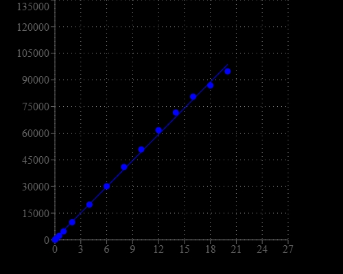
DNA Quantitation is a very important task in DNA sample preparations for various analyses. Helixyte™ Green Fluorimetric dsDNA Quantitation Kit provides a rapid method to quantify dsDNA with Helixyte™ Green BR . The assay is linear over three orders of magnitude and is a few magnitudes more sensitive than UV absorbance readings. Helixyte™ Green BR exhibits large fluorescence enhancement upon binding to dsDNA and has little sequence dependence, allowing to the accurate measurement of DNA samples from various sources, including genomic DNA, viral DNA, miniprep DNA or PCR amplification products. The assay is highly selective for double-stranded DNA (dsDNA) over RNA and is optimized to measure DNA concentrations from 10 pg/µL to 10 ng/µL.

Platform


Fluorescence microplate reader

Excitation490 nm
Emission530 nm
Cutoff515 nm
Recommended plateSolid black

Components


Example protocol


AT A GLANCE

Protocol summary

  1. Prepare dSDNA standards, test samples and dye working solution
  2. Add DNA standards or test samples (50 uL)
  3. Add Helixyte™ Green BR working solution (50 uL)
  4. Incubate at room temperature for 2 minutes
  5. Monitor fluorescence intensity at Ex/Em= 490/530 nm

Important notes
The following protocol is provided as an example for quantifying dsDNA with Helixyte™ Green BR. Warm all the components to room temperature before opening. No data are available for the mutagenicity or toxicity of Helixyte™ Green dsDNA stain. Because this reagent binds to nucleic acids, it should be treated as a potential mutagen and handled with appropriate care. The DMSO stock solution should be handled with particular caution as DMSO is known to facilitate the entry of organic molecules into tissues.

PREPARATION OF STANDARD SOLUTION

dsDNA standard

For convenience, use the Serial Dilution Planner: https://www.aatbio.com/tools/serial-dilution/17645

Add 100 uL of 100 ug/mL dsDNA Standard Solution (Component C) to 400 uL Assay Buffer (Component B) to generate 20 ug/mL dsDNA standard solution. Then perform 1:3 serial dilutions by Assay Buffer (Component B) to get serially diluted dsDNA standards ranging from 0 to 20 ug/mL.

PREPARATION OF WORKING SOLUTION

Helixyte™ Green BR working solution:
Add 50 uL Helixyte™ Green BR (Component A) into 5 mL of Assay Buffer (Component B) to make a total volume of 5.050 mL. Protect the working solution from light by covering it with foil or placing it in the dark. Note: We recommend preparing this solution in a plastic container rather than glass, as the dye may adsorb to glass surfaces. For best results, this solution should be used within a few hours of its preparation.

SAMPLE EXPERIMENTAL PROTOCOL

Table 1. Layout of dsDNA standards and test samples in a clear bottom 96-well microplate. DS=dsDNA standards (DS1-DS7, 20 to 0.027 ug/mL); BL=Blank Control; TS=Test Samples

BL

BL

TS

TS

DS1

DS1

DS2

DS2

DS3

DS3


DS4

DS4


DS5

DS5


DS6

DS6


DS7

DS7


 Table 2. Reagent composition for each well.

Well

Volume

Reagent

DS1-DS7

50 uL

Serial Dilutions (20 to 0.027 ug/mL)

BL

50 uL

Assay Buffer (Component B)

TS

50 uL

Test Sample


  1. Prepare dsDNA standards (DS), blank controls (BL), and test samples (TS) according to the layout provided in Tables 1 and 2. For a 384-well plate, use 25 uL of reagent per well instead of 50 uL. Note: Treat cells or tissue samples as desired.

  2. Add 50 uL of Helixyte™ Green BR dye working solution (2X) to each well of dsDNA standard, blank control, and test samples to make the total assay volume 100 uL/well. For a 384-well plate, add 25 uL ofHelixyte™ Green BR dye working solution into each well instead, for a total volume of 50 uL/well.

  3. Incubate the reaction for 2 minutes at room temperature, protected from light.

  4. Monitor the fluorescence intensity with a fluorescence plate reader at Ex/Em = 490/530 nm (cut off at 515nm).

Spectrum


Open in Advanced Spectrum Viewer
spectrum

Spectral properties

Excitation (nm)503
Emission (nm)527

Images


Citations


View all 34 citations: Citation Explorer
Enhanced articular cartilage regeneration using costal chondrocyte-derived scaffold-free tissue engineered constructs with ascorbic acid treatment
Authors: Zheng, Kaiwen and Ma, Yiyang and Chiu, Cheng and Xue, Mengxin and Zhang, Changqing and Du, Dajiang
Journal: Journal of Orthopaedic Translation (2024): 140--154
The Response of the Soil Microbiota to Long-Term Mineral and Organic Nitrogen Fertilization is Stronger in the Bulk Soil than in the Rhizosphere
Authors: Cardinale, Massimiliano and Ratering, Stefan and Sadeghi, Aitak and Pokhrel, Sushil and Honermeier, Bernd and Schnell, Sylvia
Journal: Genes (2020): 456
A new reporter design based on DNA origami nanostructures for quantification of short oligonucleotides using microbeads
Authors: Choi, Y., Schmidt, C., Tinnefeld, P., Bald, I., Rodiger, S.
Journal: Sci Rep (2019): 4769
Flat-top TIRF illumination boosts DNA-PAINT imaging and quantification
Authors: Stehr, F., Stein, J., Schueder, F., Schwille, P., Jungmann, R.
Journal: Nat Commun (2019): 1268
Effects of Quantification Methods, Isolation Kits, Plasma Biobanking, and Hemolysis on Cell-Free DNA Analysis in Plasma
Authors: Streleckiene, G., Forster, M., Inciuraite, R., Lukosevicius, R., Skieceviciene, J.
Journal: Biopreserv Biobank (2019): ersion="1.0" encoding="UTF-8" ?>17645.enlEndN
A universal fluorescence-based toolkit for real-time quantification of DNA and RNA nuclease activity
Authors: Sheppard, E. C., Rogers, S., Harmer, N. J., Chahwan, R.
Journal: Sci Rep (2019): 8853
Molecular-Recognition-Based DNA Nanodevices for Enhancing the Direct Visualization and Quantification of Single Vesicles of Tumor Exosomes in Plasma Microsamples
Authors: He, D., Ho, S. L., Chan, H. N., Wang, H., Hai, L., He, X., Wang, K., Li, H. W.
Journal: Anal Chem (2019): 2768-2775
Quantification of fixed adherent cells using a strong enhancer of the fluorescence of DNA dyes
Authors: Ligasova, A., Koberna, K.
Journal: Sci Rep (2019): 8701
Identification and Quantification of Heterogeneously-methylated DNA Fragments Using Epiallele-sensitive Droplet Digital Polymerase Chain Reaction (EAST-ddPCR)
Authors: Menschikowski, M., J and eck, C., Friedemann, M., Richter, S., Thiem, D., Lange, B. S., Suttorp, M.
Journal: Cancer Genomics Proteomics (2018): 299-312
A fluorescent reporter for quantification and enrichment of DNA editing by APOBEC-Cas9 or cleavage by Cas9 in living cells
Authors: St Martin, A., Salamango, D., Serebrenik, A., Shaban, N., Brown, W. L., Donati, F., Munagala, U., Conticello, S. G., Harris, R. S.
Journal: Nucleic Acids Res (2018): e84