logo
AAT Bioquest

Helixyte™ Green Nucleic Acid Gel Stain *10,000X DMSO Solution*

Helixyte™ Green is manufactured by AAT Bioquest, and it has the same chemical structure of SYBR® Green (SYBR® is the trademark of ThermoFisher). Helixyte™ Green is an excellent nucleic acid gel stain. It has the same spectral properties to those of SYBR® Green, thus a great replacement to SYBR® Green (SYBR® Green is the trademark of ThermoFisher). It is one of the most sensitive stains available for detecting double-stranded DNA (dsDNA) in agarose and polyacrylamide gels. Helixyte™ Green has much greater sensitivity for dsDNA, thus especially useful for assays where the presence of contaminating RNA or ssDNA might obscure results. Helixyte™ Green stain is ideal for use with laser scanners with the same instrument settings of SYBR Green. Helixyte™ Green is much more sensitive than ethidium bromide for DNA in agarose gels. The gels soaked in diluted Helixyte™ Green stain can be visualized without desalting. It is compatible with UV transilluminators, gel documentation systems, and laser scanners.
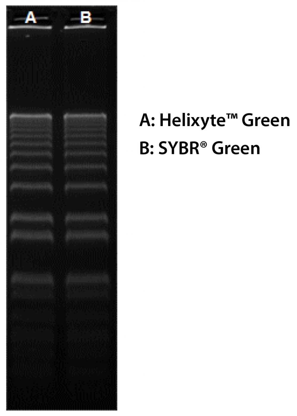
Example protocol

AT A GLANCE

Spectral Properties of Helixyte™ Green Nucleic Acid Gel Stain

Excitation/Emission: 497/521 nm when bound to DNA

Important Note

Helixyte™ Green nucleic acid gel stain is significantly less mutagenic than ethidium bromide. Nevertheless, we must caution that there is currently no available data on the mutagenicity or toxicity of Helixyte™ Green stain in humans. It is prudent to treat this reagent as a potential mutagen and handle it with appropriate care, especially considering its binding affinity to nucleic acids. Additionally, ensure proper disposal of the stain in compliance with local regulations.

We have observed that the greatest sensitivity is attained through post-staining, which eliminates the risk of dye interference with DNA migration. While the precast protocol is more convenient, some DNA samples may experience migration issues. Hence, it is strongly advised to limit the gel running time to no more than 2 hours. The provided protocols are recommendations, and conducting comparisons may be necessary to determine which one better meets your specific needs.

PREPARATION OF WORKING SOLUTION

Helixyte™ Green Working Wolution (1X)
  1. Prepare a 1X Helixyte™ Green working solution by diluting the 10,000X stock reagent with a pH 7.5 - 8 buffer, such as TAE, TBE, or TE. Buffers with a pH of 8.0 are preferred.

    Note: Staining solutions prepared in water are less stable than those prepared in buffer and must be used within 24 hours to ensure maximal staining sensitivity.

    Note: In addition, staining solutions prepared in buffers with pH below 7.5 or above 8.0 are less stable and show reduced staining efficacy.

SAMPLE EXPERIMENTAL PROTOCOL

Post-Staining Protocol
  1. Run gels based on your standard protocol.

  2. Place the gel in a suitable polypropylene container. Gently add a sufficient amount of the 1X Helixyte™ Green working solution to submerge the gel.

    Note: Do not use a glass container, as it will adsorb much of the dye in the staining solution.

  3. Agitate the gel gently at room temperature for ~30 minutes, protected from the light.

    Note: The staining solution can be stored in the dark (preferably refrigerated) for a week and reused up to 2 - 3 times.

  4. Image the stained gel with a 254 nm transilluminator or a laser-based gel scanner using a long path green filter, such as a SYBR® filter or GelStar® filter.

Pre-Casting Protocol
  1. Prepare agarose gel solution using your standard protocol.

  2. Add 1X Helixyte™ Green working solution to the gel and mix thoroughly.

  3. Run gels based on your standard protocol.

  4. Image the stained gel with a 254 nm transilluminator or a laser-based gel scanner using a long path green filter, such as a SYBR® filter or GelStar® filter.

DNA-Staining Before Electrophoresis
  1. Incubate DNA with a 1:3000 to 1:10,000 dilution of the dye (in TE, TBE, or TAE) for at least 15 minutes prior to electrophoresis.

  2. Run gels based on your standard protocol.

  3. Image the stained gel with a 254 nm transilluminator, or a laser-based gel scanner using a long path green filter such as a SYBR® filter or GelStar® filter.

Spectrum

Product family

Citations

View all 2 citations: Citation Explorer
A promiscuous DNA packaging machine from bacteriophage T4
Authors: Zhang, Zhihong and Kottadiel, Vishal I and Vafabakhsh, Reza and Dai, Li and Chemla, Yann R and Ha, Taekjip and Rao, Venigalla B
Journal: PLoS biology (2011): e1000592

References

View all 22 references: Citation Explorer
Plasmalemma permeability and necrotic cell death phenotypes after intracerebral hemorrhage in mice
Authors: Zhu X, Tao L, Tejima-M and eville E, Qiu J, Park J, Garber K, Ericsson M, Lo EH, Whalen MJ.
Journal: Stroke (2012): 524
Intra-organ Biodistribution of Gold Nanoparticles Using Intrinsic Two-photon Induced Photoluminescence
Authors: Park J, Estrada A, Schwartz JA, Diagaradjane P, Krishnan S, Dunn AK, Tunnell JW.
Journal: Lasers Surg Med (2010): 630
Screening by imaging: scaling up single-DNA-molecule analysis with a novel parabolic VA-TIRF reflector and noise-reduction techniques
Authors: van 't Hoff M, Reuter M, Dryden DT, Oheim M.
Journal: Phys Chem Chem Phys (2009): 7713
Novel anatomic structures in the brain and spinal cord of rabbit that may belong to the Bonghan system of potential acupuncture meridians
Authors: Lee BC, Kim S, Soh KS.
Journal: J Acupunct Meridian Stud (2008): 29
Triplet fraction buildup effect of the DNA-YOYO complex studied with fluorescence correlation spectroscopy
Authors: Shimizu M, Sasaki S, Kinjo M.
Journal: Anal Biochem (2007): 87
Page updated on December 11, 2024

Ordering information

Price
Unit size
1 ml
100 ul
Catalog Number
1759017604
Quantity
Add to cart

Additional ordering information

Telephone1-800-990-8053
Fax1-800-609-2943
Emailsales@aatbio.com
InternationalSee distributors
Bulk requestInquire
Custom sizeInquire
Technical SupportContact us
Purchase orderSend to sales@aatbio.com
ShippingStandard overnight for United States, inquire for international
Request quotation

Physical properties

Molecular weight

N/A

Solvent

DMSO

Spectral properties

Excitation (nm)

498

Emission (nm)

522

Storage, safety and handling

H-phraseH303, H313, H340
Hazard symbolT
Intended useResearch Use Only (RUO)
R-phraseR20, R21, R68

Storage

Freeze (< -15 °C); Minimize light exposure
UNSPSC41116134