Products
Services
Resources
Selection Guides
About
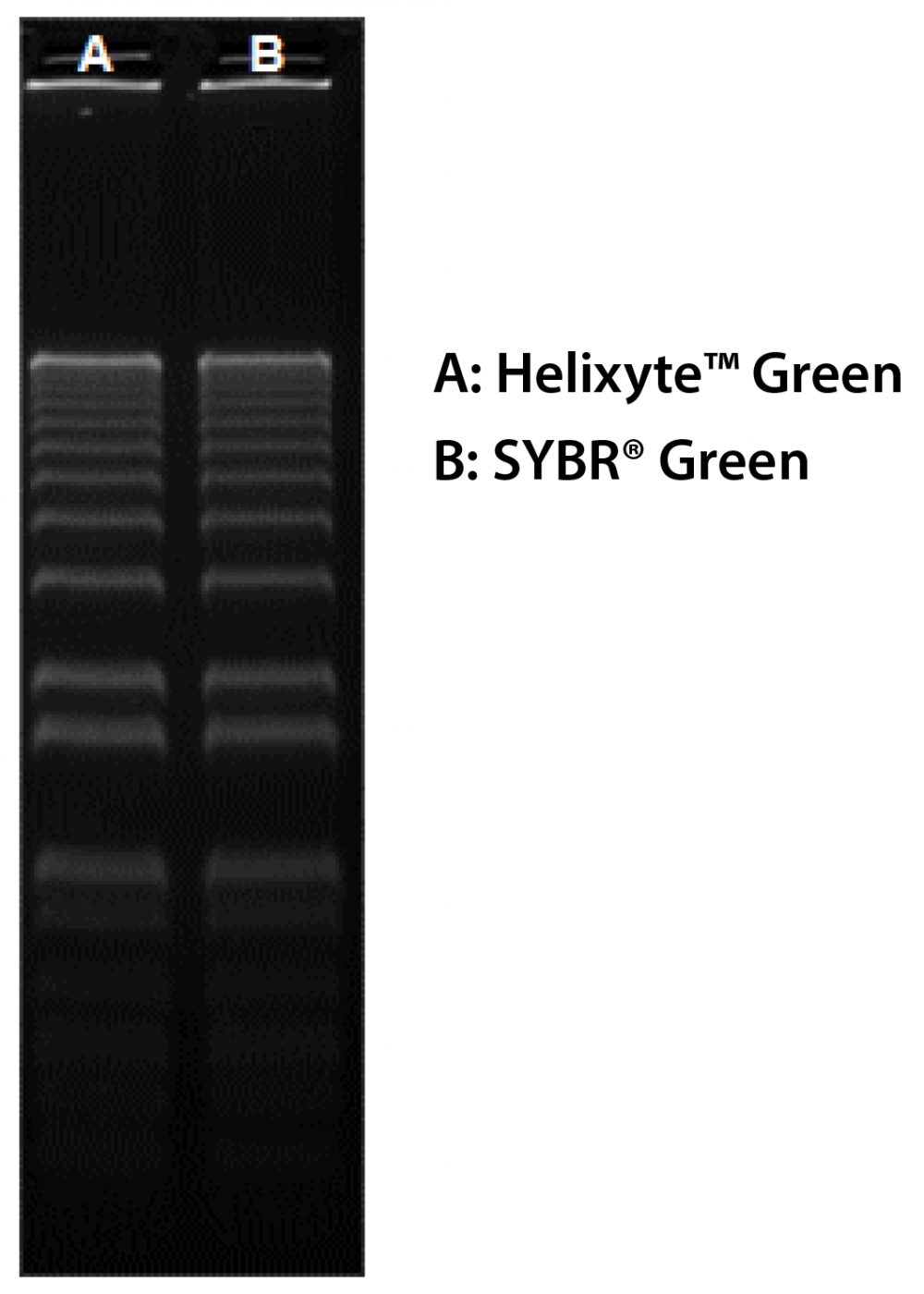
Helixyte™ Green Nucleic Acid Gel Stain
10,000X DMSO Solution
Helixyte™ Green is manufactured by AAT Bioquest, and it has the same chemical structure of SYBR® Green (SYBR® is the trademark of ThermoFisher). Helixyte™ Green is an excellent nucleic acid gel stain. It has the same spectral properties to those of SYBR® Green, thus a great replacement to SYBR® Green (SYBR® Green is the trademark of ThermoFisher). It is one of the most sensitive stains available for detecting double-stranded DNA (dsDNA) in agarose and polyacrylamide gels. Helixyte™ Green has much greater sensitivity for dsDNA, thus especially useful for assays where the presence of contaminating RNA or ssDNA might obscure results. Helixyte™ Green stain is ideal for use with laser scanners with the same instrument settings of SYBR Green. Helixyte™ Green is much more sensitive than ethidium bromide for DNA in agarose gels. The gels soaked in diluted Helixyte™ Green stain can be visualized without desalting. It is compatible with UV transilluminators, gel documentation systems, and laser scanners.
| Catalog | Size | Price | Quantity |
|---|---|---|---|
| 17590 | 1 ml | Price | |
| 17604 | 100 ul | Price |

Physical properties
| Molecular weight | N/A |
| Solvent | DMSO |
Spectral properties
| Excitation (nm) | 498 |
| Emission (nm) | 522 |
Usage and storage
| Intended use | Research Use Only (RUO) |
| Storage | Freeze (< -15 °C); Minimize light exposure |
Contact us
| Telephone | |
| Fax | |
| sales@aatbio.com | |
| International | See distributors |
| Bulk request | Inquire |
| Custom size | Inquire |
| Technical Support | Contact us |
| Request quotation | Request |
| Purchase order | Send to sales@aatbio.com |
| Shipping | Standard overnight for United States, inquire for international |
Page updated on July 28, 2026