logo
AAT Bioquest

mFluor™ Green 620 SE

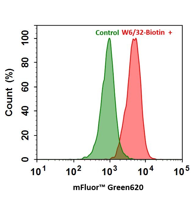
mFluor™ Green 620 dye is a unique dye that is well excited by the green laser at 532 nm to give red fluorescence. mFluor™ Green 620 dye is water-soluble, and the protein conjugates prepared with mFluor™ Green 620 dye are bright with a Stokes Shift of ~80 nm. mFluor™ Green 620 dye and conjugates are excellent green laser reagents for both flow cytometry research and fluorescence imaging applications.

Example protocol

PREPARATION OF STOCK SOLUTIONS

Unless otherwise noted, all unused stock solutions should be divided into single-use aliquots and stored at -20 °C after preparation. Avoid repeated freeze-thaw cycles.

1. Protein stock solution (Solution A)
Mix 100 µL of a reaction buffer (e.g., 1 M  sodium carbonate solution or 1 M phosphate buffer with pH ~9.0) with 900 µL of the target protein solution (e.g. antibody, protein concentration >2 mg/mL if possible) to give 1 mL protein labeling stock solution.
Note     The pH of the protein solution (Solution A) should be 8.5 ± 0.5. If the pH of the protein solution is lower than 8.0, adjust the pH to the range of 8.0-9.0 using 1 M  sodium bicarbonate solution or 1 M pH 9.0 phosphate buffer.
Note     The protein should be dissolved in 1X phosphate buffered saline (PBS), pH 7.2-7.4. If the protein is dissolved in Tris or glycine buffer, it must be dialyzed against 1X PBS, pH 7.2-7.4, to remove free amines or ammonium salts (such as ammonium sulfate and ammonium acetate) that are widely used for protein precipitation.
Note     Impure antibodies or antibodies stabilized with bovine serum albumin (BSA) or gelatin will not be labeled well. The presence of sodium azide or thimerosal might also interfere with the conjugation reaction. Sodium azide or thimerosal can be removed by dialysis or spin column for optimal labeling results.
Note     The conjugation efficiency is significantly reduced if the protein concentration is less than 2 mg/mL. For optimal labeling efficiency the final protein concentration range of 2-10 mg/mL is recommended.


2. mFluor™ Green 620 SE stock solution (Solution B)
Add anhydrous DMSO into the vial of mFluor™ Green 620 SE to make a 10 mM stock solution. Mix well by pipetting or vortex.
Note     Prepare the dye stock solution (Solution B) before starting the conjugation. Use promptly. Extended storage of the dye stock solution may reduce the dye activity. Solution B can be stored in freezer for two weeks when kept from light and moisture. Avoid freeze-thaw cycles.

SAMPLE EXPERIMENTAL PROTOCOL

This labeling protocol was developed for the conjugate of Goat anti-mouse IgG with mFluor™ Green 620 SE. You might need further optimization for your particular proteins.
Note     Each protein requires distinct dye/protein ratio, which also depends on the properties of dyes. Over labeling of a protein could detrimentally affects its binding affinity while the protein conjugates of low dye/protein ratio gives reduced sensitivity.


Run conjugation reaction
  1. Use 10:1 molar ratio of Solution B (dye)/Solution A (protein) as the starting point:  Add 5 µL of the dye stock solution (Solution B, assuming the dye stock solution is 10 mM) into the vial of the protein solution (95 µL of Solution A) with effective shaking. The concentration of the protein is ~0.05 mM assuming the protein concentration is 10 mg/mL and the molecular weight of the protein is ~200KD.
    Note     We recommend to use 10:1 molar ratio of Solution B (dye)/Solution A (protein). If it is too less or too high, determine the optimal dye/protein ratio at 5:1, 15:1 and 20:1 respectively.
  2. Continue to rotate or shake the reaction mixture at room temperature for 30-60 minutes. 

Purify the conjugation
The following protocol is an example of dye-protein conjugate purification by using a Sephadex G-25 column.
  1. Prepare Sephadex G-25 column according to the manufacture instruction.
  2. Load the reaction mixture (From "Run conjugation reaction") to the top of the Sephadex G-25 column.
  3. Add PBS (pH 7.2-7.4) as soon as the sample runs just below the top resin surface.
  4. Add more PBS (pH 7.2-7.4) to the desired sample to complete the column purification. Combine the fractions that contain the desired dye-protein conjugate.
    Note     For immediate use, the dye-protein conjugate need be diluted with staining buffer, and aliquoted for multiple uses.
    Note     For longer term storage, dye-protein conjugate solution need be concentrated or freeze dried. 

Calculators

Common stock solution preparation

Table 1. Volume of DMSO needed to reconstitute specific mass of mFluor™ Green 620 SE to given concentration. Note that volume is only for preparing stock solution. Refer to sample experimental protocol for appropriate experimental/physiological buffers.

0.1 mg0.5 mg1 mg5 mg10 mg
1 mM113.125 µL565.624 µL1.131 mL5.656 mL11.312 mL
5 mM22.625 µL113.125 µL226.249 µL1.131 mL2.262 mL
10 mM11.312 µL56.562 µL113.125 µL565.624 µL1.131 mL

Molarity calculator

Enter any two values (mass, volume, concentration) to calculate the third.

Mass (Calculate)Molecular weightVolume (Calculate)Concentration (Calculate)Moles
/=x=

Spectrum

Product family

NameExcitation (nm)Emission (nm)Extinction coefficient (cm -1 M -1)Quantum yieldCorrection Factor (260 nm)Correction Factor (280 nm)
mFluor™ Blue 620 SE5896169800010.1810.6830.849

Citations

View all 3 citations: Citation Explorer
Deep Sequencing Analysis of the Eha-Regulated Transcriptome of Edwardsiella tarda Following Acidification
Authors: Gao, D and Liu, N and Li, Y and Zhang, Y and Liu, G and others, undefined
Journal: Metabolomics (Los Angel) (2017): 2153--0769
Suramin inhibits cullin-RING E3 ubiquitin ligases
Authors: Wu, Kenneth and Chong, Robert A and Yu, Qing and Bai, Jin and Spratt, Donald E and Ching, Kevin and Lee, Chan and Miao, Haibin and Tappin, Inger and Hurwitz, Jerard and others, undefined
Journal: Proceedings of the National Academy of Sciences (2016): E2011--E2018
Glycosaminoglycan mimicry by COAM reduces melanoma growth through chemokine induction and function
Authors: Piccard, Helene and Berghmans, Nele and Korpos, Eva and Dillen, Chris and Aelst, Ilse Van and Li, S and ra , undefined and Martens, Erik and Liekens, S and ra , undefined and Noppen, Sam and Damme, Jo Van and others, undefined
Journal: International Journal of Cancer (2012): E425--E436

References

View all 49 references: Citation Explorer
Sequential ordering among multicolor fluorophores for protein labeling facility via aggregation-elimination based beta-lactam probes
Authors: Sadhu KK, Mizukami S, Watanabe S, Kikuchi K.
Journal: Mol Biosyst (2011): 1766
Visualizing dengue virus through Alexa Fluor labeling
Authors: Zhang S, Tan HC, Ooi EE.
Journal: J Vis Exp. (2011)
Fluorescent "Turn-on" system utilizing a quencher-conjugated peptide for specific protein labeling of living cells
Authors: Arai S, Yoon SI, Murata A, Takabayashi M, Wu X, Lu Y, Takeoka S, Ozaki M.
Journal: Biochem Biophys Res Commun (2011): 211
Neuroanatomical basis of clinical joint application of "Jinggu" (BL 64, a source-acupoint) and "Dazhong" (KI 4, a Luo-acupoint) in the rat: a double-labeling study of cholera toxin subunit B conjugated with Alexa Fluor 488 and 594
Authors: Cui JJ, Zhu XL, Ji CF, Jing XH, Bai WZ.
Journal: Zhen Ci Yan Jiu (2011): 262
Simultaneous detection of virulence factors from a colony in diarrheagenic Escherichia coli by a multiplex PCR assay with Alexa Fluor-labeled primers
Authors: Kuwayama M, Shigemoto N, Oohara S, Tanizawa Y, Yamada H, Takeda Y, Matsuo T, Fukuda S.
Journal: J Microbiol Methods (2011): 119
Page updated on September 18, 2024

Ordering information

Price
Unit size
Catalog Number1165
Quantity
Add to cart

Additional ordering information

Telephone1-800-990-8053
Fax1-800-609-2943
Emailsales@aatbio.com
InternationalSee distributors
Bulk requestInquire
Custom sizeInquire
Technical SupportContact us
Purchase orderSend to sales@aatbio.com
ShippingStandard overnight for United States, inquire for international
Request quotation

Physical properties

Molecular weight

883.98

Solvent

DMSO

Spectral properties

Absorbance (nm)

525

Correction Factor (260 nm)

0.895

Correction Factor (280 nm)

0.569

Extinction coefficient (cm -1 M -1)

500001

Excitation (nm)

525

Emission (nm)

623

Storage, safety and handling

H-phraseH303, H313, H333
Hazard symbolXN
Intended useResearch Use Only (RUO)
R-phraseR20, R21, R22

Storage

Freeze (< -15 °C); Minimize light exposure
UNSPSC12171501
Flow cytometry analysis of HL-60 cells stained with (Red) or without (Green) 1ug/ml Anti-Human HLA-ABC-Biotin and then followed by mFluor&trade; Green 620-streptavidin conjugate.
Spectral signature of mFluor™ Green 620 dye. Data acquired on a 4-laser Cytek Aurora and normal human peripheral blood cells stained with clone SK3 (CD4) conjugated to mFluor™ Green 620 dye (Cat. No. 100420U0) were used for analysis.

Alternative formats