logo
AAT Bioquest

Screen Quest™ Calbryte-590 Probenecid-Free and Wash-Free Calcium Assay Kit

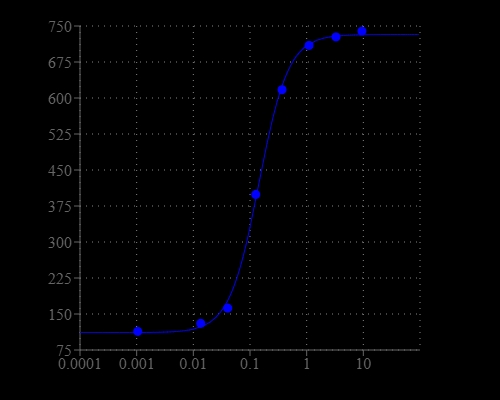
Graph illustrates signal-to-noise ratio (SNR) x 100%. ATP dose response was measured in CHO-K1 cells with Screen Quest™ Calbryte 590 Probenecid-Free and Wash-Free Calcium Assay Kit. CHO-K1 cells were seeded overnight at 50,000 cells/100 µL/well in a 96-well black wall/clear bottom costar plate. 100 µL dye loading solution was added and incubated for 45 min at 37°C and 15 min at RT. ATP (50 µL/well) was added by FlexStation 3 to achieve the final indicated concentrations.
Ordering information
Price
Catalog Number
Unit Size
Quantity
Add to cart
Additional ordering information
Telephone1-800-990-8053
Fax1-800-609-2943
Emailsales@aatbio.com
InternationalSee distributors
Bulk requestInquire
Custom sizeInquire
ShippingStandard overnight for United States, inquire for international
Request quotation
Spectral properties
Excitation (nm)581
Emission (nm)593
Storage, safety and handling
H-phraseH303, H313, H333
Hazard symbolXN
Intended useResearch Use Only (RUO)
R-phraseR20, R21, R22
UNSPSC12352200
Related products
Screen Quest™ Membrane Potential Assay Kit *Orange Fluorescence*
Screen Quest™ Membrane Potential Assay Kit *Red Fluorescence*
Screen Quest™ 10X cell staining buffer with Phenol Red Plus™
Screen Quest™ 10X calcium assay buffer with Phenol Red Plus™
Screen Quest™ Luminometric Calcium Assay Kit
Screen Quest™ Fluo-8 Medium Removal Calcium Assay Kit *Optimized for Difficult Cell Lines*
Screen Quest™ Fluo-8 No Wash Calcium Assay Kit
Screen Quest™ Fura-2 No Wash Calcium Assay Kit
Screen Quest™ Rhod-4 No Wash Calcium Assay Kit *Medium Removal*
Screen Quest™ Rhod-4 No Wash Calcium Assay Kit
Screen Quest™ Fluorimetric MDR Assay Kit
Screen Quest™ Colorimetric Chloride Channel Assay Kit
Screen Quest™ Colorimetric ELISA cAMP Assay Kit
Screen Quest™ Fluorimetric ELISA cAMP Assay Kit
Screen Quest™ TR-FRET No Wash cAMP Assay Kit
Screen Quest™ Live Cell cAMP Assay Service Pack
Screen Quest™ Fluorimetric Fatty Acid Uptake Assay Kit
Screen Quest™ Fluorimetric Glucose Uptake Assay Kit
Screen Quest™ Colorimetric Glucose Uptake Assay Kit
Screen Quest™ HEK-CNGC-Amylin 3 Receptor Cells
Screen Quest™ HEK-CNGC-Cannabinoid Receptor 1 Cells
Screen Quest™ CHO-Ga16- Chemokine (C-C) Receptor 2B Cells
Screen Quest™ HEK-CNGC-Chemokine (C-X-C motif) Receptor 4 Cells
Screen Quest™ HEK-CNGC-Dopamine Receptor 1 (DRD1) Cells
Screen Quest™ HEK-CNGC-Glucagon-like Receptor 1 (GLP1R) Cells
Screen Quest™ HEK-CNGC-Opiate Receptor-like 1 (ORL1) Cells
Screen Quest™ Human Nociceptin Receptor Ga16 coupled CHO Cells (NOP-Ga16)
Screen Quest™ CHO-Gqi Chimera Cell line
Screen Quest™ CHO-Gqo Chimera Cell line
Screen Quest™ CHO-Gqz Chimera Cell line
Screen Quest™ CHO-Gqs Chimera Cell line
Screen Quest™ CHO-Ga16 Chimera Cell line
Screen Quest™ Fluo-4 No Wash Calcium Assay Kit
Screen Quest™ No Wash Potassium Channel Assay Kit
Chemical Phosphorylation Reagent I (CPR I)
Cell Meter™ Mitochondrial Hydroxyl Radical Detection Kit *Red Fluorescence*
Cal Green™ 1, hexapotassium salt
Cal Green™ 1, AM [Equivalent to Calcium Green-1, AM]
Cal-590™-Dextran Conjugate *MW 3,000*
Cal-590™-Dextran Conjugate *MW 10,000*
Cal-590™ AM
Cal-590™, sodium salt
Cal-590™, potassium salt
Cal-630™ AM
Cal-630™, sodium salt
Cal-630™, potassium salt
Cal-630™-Dextran Conjugate *MW 3,000*
Cal-630™-Dextran Conjugate *MW 10,000*
Fluo-4 AM *Ultrapure Grade* *CAS 273221-67-3*
Fluo-4, Pentapotassium Salt
Cal Red™ R525/650 potassium salt
Cal Red™ R525/650 AM
Cal-520®-Dextran Conjugate *MW 3,000*
Cal-520®-Dextran Conjugate *MW 10,000*
Cal-520®-Biotin Conjugate
Cal-520®-Biocytin Conjugate
Cal-520® NHS Ester
Cal-520® maleimide
Fluo-3, AM *CAS 121714-22-5*
Fluo-3, AM *UltraPure grade* *CAS 121714-22-5*
Fluo-3, AM *Bulk package* *CAS 121714-22-5*
Fluo-3FF, AM *UltraPure grade* *Cell permeant*
Fluo-3, pentasodium salt
Fluo-3, pentapotassium salt
Fluo-3, pentaammonium salt
Fluo-3FF, pentapotassium salt
Fluo-8®, AM
Fluo-8®, sodium salt
Fluo-8®, potassium salt
Fluo-8H™, AM
Fluo-8H™, sodium salt
Fluo-8L™, AM
Fluo-8L™, sodium salt
Fluo-8L™, potassium salt
Fluo-8FF™, potassium salt
Fluo-8FF™, AM
Cal-520®, AM
Cal-520®, sodium salt
Cal-520®, potassium salt
Cal-520FF™, AM
Cal-520FF™, potassium salt
Mag-Fluo-4 potassium salt
Mag-Fluo-4 AM
Fluo-2, potassium salt
Fluo-2, AM
Fluo-5F, AM *Cell permeant*
Fluo-5F, pentapotassium Salt *Cell impermeant*
Fluo-5N, AM *Cell permeant*
Fluo-5N, pentapotassium Salt *Cell impermeant*
Cal-520N™, AM
Cal-520N™, potassium salt
Calbryte™ 520 AM
Calbryte™ 520, potassium salt
Calbryte™ 590 AM
Calbryte™ 590, potassium salt
Calbryte™ 630 AM
Calbryte™ 630, potassium salt
Calbryte™-520L AM
Calbryte™-520L, potassium salt
Cal-500™, potassium salt
Cal-500™ AM
Cal-670™, potassium salt
Cal-670™-Dextran Conjugate *MW 3,000*
Cal-670™-Dextran Conjugate *MW 10,000*
Cal-770™, potassium salt
Cal-770™-Dextran Conjugate *MW 3,000*
Cal-770™-Dextran Conjugate *MW 10,000*
Calbryte™-520XL azide
Calbryte™-520XL, potassium salt
Calbryte™-520XL AM
Calbryte™-520XL-Dextran
Cal-520L®-Dextran Conjugate *MW 10,000*
RatioWorks™ Cal-520L®/Cy5-Dextran Conjugate *MW 10,000*
Cal-520L™ maleimide
RatioWorks™ Cal-520®/zFluor 647™ -Dextran Conjugate *MW 10,000*
Cal-590L® Dextran Conjugate *MW 10,000*
RatioWorks™ Cal-590L®/Cy5-Dextran Conjugate *MW 10,000*
Cal-520® amine
Cal-520® azide
Cal-520® alkyne
Show More (110)

OverviewpdfSDSpdfProtocol


Excitation (nm)
581
Emission (nm)
593
Calcium flux assays are the preferred methods for screening G protein coupled receptors (GPCRs) in drug discovery. Screen Quest™ Calbryte-590™ Probenecid-Free and Wash-Free Calcium Assay Kit provides the most robust homogeneous red fluorescence-based assay for detecting the intracellular calcium mobilization. Cells expressing a GPCR of interest that signals through calcium are pre-loaded with our proprietary Calbryte-590 AM which can cross cell membrane. Calbryte-590 AM is the brightest calcium indicator available for HTS screening. Once inside the cell, the lipophilic blocking groups of Calbryte-590 AM are cleaved by non-specific cell esterase, resulting in a negatively charged fluorescent dye that stays inside cells, and its fluorescence is greatly enhanced upon binding to calcium. When cells stimulated with screening compounds, the receptor signals release of intracellular calcium, which greatly increase the fluorescence of Calbryte-590 AM. The characteristics of its excellent cell retention, high sensitivity, and 100-250 times fluorescence increases (when it forms complexes with calcium) make Calbryte-590 AM an ideal indicator for measurement of cellular calcium. Calbryte-590 AM is the only red calcium dye that does not require probenecid to improve cellular retention. This Screen Quest™ Calbryte-590™ Probenecid-Free and Wash-Free Calcium Assay Kit provides the most optimized assay method for monitoring GPCRs and calcium channels with fragile or difficult cell lines. Compared to the green fluorescence-based calcium assays, this assay kit has less interference from the colored compounds from a compound library. It is also compatible with GFP cell lines for high content analysis. The assay can be performed in a convenient 96-well or 384-well microtiter-plate format and easily adapted to automation.
Platform


Fluorescence microplate reader

Excitation540 nm
Emission590 nm
Cutoff570 nm
Recommended plateBlack wall/clear bottom
Instrument specification(s)Bottom read mode/programmable liquid handling

Other instruments

ArrayScan, FDSS, FlexStation, IN Cell Analyzer, NOVOStar, ViewLux

Components


Example protocol


AT A GLANCE

Protocol summary

  1. Prepare cells in growth medium
  2. Add Calbryte™ 590 AM dye-loading solution (100 µL/well for 96-well plate or 25 µL/well for 384-well plate)
  3. Incubate at room temperature or 37°C for 45-60 minutes
  4. Monitor fluorescence at Ex/Em = 540/590 nm

Important notes
Thaw all the kit components at room temperature before use.

PREPARATION OF STOCK SOLUTION

Unless otherwise noted, all unused stock solutions should be divided into single-use aliquots and stored at -20 °C after preparation. Avoid repeated freeze-thaw cycles.

1. Calbryte™ 590 AM stock solution:
Add 20 µL (Cat. # 36200) or 200 µL (Cat. # 36201 and # 36202) of DMSO into the vial of Calbryte™ 590 AM (Component A) and mix them well. Note: 20 µL of Calbryte™ 590 AM stock solution is enough for one plate. Unused Calbryte™ 590 AM stock solution can be aliquoted and stored at < -20 oC for more than one month if the tubes are sealed tightly. Note: Protect from light and avoid repeated freeze-thaw cycles.

2. Assay buffer (1X):
Mix 9 mL of HHBS (Component C, not included in the kit Cat. # 36202) with 1 mL of 10X Pluronic® F127 Plus (10X) (Component B) and mix them well.

PREPARATION OF WORKING SOLUTION

Calbryte™ 590 AM dye-loading solution:
Add 20 µL of Calbryte™ 590 AM stock solution into 10 mL of Assay Buffer (1X) and mix them well. Note: This working solution is stable for at least 2 hours at room temperature. Note: 10 mL dye-loading solution is enough for one 96-wells plate.

For guidelines on cell sample preparation, please visit
https://www.aatbio.com/resources/guides/cell-sample-preparation.html

SAMPLE EXPERIMENTAL PROTOCOL

  1. Add 100 µL/well (96-well plate) or 25 µL/well (384-well plate) of Calbryte™ 590 AM dye-loading solution into the cell plate.

  2. Incubate the dye-loading plate in a cell incubator for 60 minutes, and then incubate the plate at room temperature for another 15 - 30 minutes. Note: If the assay requires 37°C, perform the experiment immediately without further room temperature incubation. If the cells can function well at room temperature for longer time, incubate the cell plate at room temperature for 1 hour (It is recommended that the incubation time be no longer than 2 hours.)

  3. Prepare the compound plate with HHBS or your desired buffer.

  4. Run the calcium flux assay by monitoring the fluorescence intensity at Ex/Em = 540/590 nm.

Spectrum


Open in Advanced Spectrum Viewer
spectrum

Spectral properties

Excitation (nm)581
Emission (nm)593

Product Family


NameExcitation (nm)Emission (nm)Quantum yield
Screen Quest™ Calbryte-520 Probenecid-Free and Wash-Free Calcium Assay Kit4935150.751

Images


Citations


View all 10 citations: Citation Explorer
Calreticulin regulates TGF-&beta;1-induced epithelial mesenchymal transition through modulating Smad signaling and calcium signaling
Authors: Wu, Yanjiao and Xu, Xiaoli and Ma, Lunkun and Yi, Qian and Sun, Weichao and Tang, Liling
Journal: The International Journal of Biochemistry &amp; Cell Biology (2017)
Monosialoganglioside 1 may alleviate neurotoxicity induced by propofol combined with remifentanil in neural stem cells
Authors: Lu, Jiang and Yao, Xue-qin and Luo, Xin and Wang, Yu and Chung, Sookja Kim and Tang, He-xin and Cheung, Chi Wai and Wang, Xian-yu and Meng, Chen and Li, Qing and others, undefined
Journal: Neural Regeneration Research (2017): 945
Obtaining spontaneously beating cardiomyocyte-like cells from adipose-derived stromal vascular fractions cultured on enzyme-crosslinked gelatin hydrogels
Authors: Yang, Gang and Xiao, Zhenghua and Ren, Xiaomei and Long, Haiyan and Ma, Kunlong and Qian, Hong and Guo, Yingqiang
Journal: Scientific Reports (2017): 41781
Dexmedetomidine reduces hypoxia/reoxygenation injury by regulating mitochondrial fission in rat hippocampal neurons
Authors: Liu, Jia and Du, Qing and Zhu, He and Li, Yu and Liu, Maodong and Yu, Shoushui and Wang, Shilei
Journal: Int J Clin Exp Med (2017): 6861--6868
Di (2-ethylhexyl) phthalate-induced apoptosis in rat INS-1 cells is dependent on activation of endoplasmic reticulum stress and suppression of antioxidant protection
Authors: Sun, Xia and Lin, Yi and Huang, Qiansheng and Shi, Junpeng and Qiu, Ling and Kang, Mei and Chen, Yajie and Fang, Chao and Ye, Ting and Dong, Sijun
Journal: Journal of cellular and molecular medicine (2015): 581--594
The effect of mitochondrial calcium uniporter on mitochondrial fission in hippocampus cells ischemia/reperfusion injury
Authors: Zhao, Lantao and Li, Shuhong and Wang, Shilei and Yu, Ning and Liu, Jia
Journal: Biochemical and biophysical research communications (2015): 537--542
Role of mitochondrial calcium uniporter in regulating mitochondrial fission in the cerebral cortexes of living rats
Authors: Liang, Nan and Wang, Peng and Wang, Shilei and Li, Shuhong and Li, Yu and Wang, Jinying and Wang, Min
Journal: Journal of Neural Transmission (2014): 593--600
Propofol and remifentanil at moderate and high concentrations affect proliferation and differentiation of neural stem/progenitor cells
Authors: Li, Qing and Lu, Jiang and Wang, Xianyu and others, undefined
Journal: Neural regeneration research (2014): 2002
Fungus induces the release of IL-8 in human corneal epithelial cells, via Dectin-1-mediated protein kinase C pathways.
Authors: Peng, Xu-Dong and Zhao, Gui-Qiu and Lin, Jing and Jiang, Nan and Xu, Qiang and Zhu, Cheng-Cheng and Qu, Jain-Qiu and Cong, Lin and Li, Hui
Journal: International journal of ophthalmology (2014): 441--447
Increased expression of cell adhesion molecule 1 by mast cells as a cause of enhanced nerve--mast cell interaction in a hapten-induced mouse model of atopic dermatitis
Authors: Hagiyama, M and Inoue, T and Furuno, T and Iino, T and Itami, S and Nakanishi, M and Asada, H and Hosokawa, Y and Ito, A
Journal: British Journal of Dermatology (2013): 771--778

References


View all 53 references: Citation Explorer
A flow cytometric comparison of Indo-1 to fluo-3 and Fura Red excited with low power lasers for detecting Ca(2+) flux
Authors: Bailey S, Macardle PJ.
Journal: J Immunol Methods (2006): 220
Functional fluo-3/AM assay on P-glycoprotein transport activity in L1210/VCR cells by confocal microscopy
Authors: Orlicky J, Sulova Z, Dovinova I, Fiala R, Zahradnikova A, Jr., Breier A.
Journal: Gen Physiol Biophys (2004): 357
Comparison of human recombinant adenosine A2B receptor function assessed by Fluo-3-AM fluorometry and microphysiometry
Authors: Patel H, Porter RH, Palmer AM, Croucher MJ.
Journal: Br J Pharmacol (2003): 671
Measurement of the dissociation constant of Fluo-3 for Ca2+ in isolated rabbit cardiomyocytes using Ca2+ wave characteristics
Authors: Loughrey CM, MacEachern KE, Cooper J, Smith GL.
Journal: Cell Calcium (2003): 1
A sensitive method for the detection of foot and mouth disease virus by in situ hybridisation using biotin-labelled oligodeoxynucleotides and tyramide signal amplification
Authors: Zhang Z, Kitching P.
Journal: J Virol Methods (2000): 187
Kinetics of onset of mouse sperm acrosome reaction induced by solubilized zona pellucida: fluorimetric determination of loss of pH gradient between acrosomal lumen and medium monitored by dapoxyl (2-aminoethyl) sulfonamide and of intracellular Ca(2+) chang
Authors: Rockwell PL, Storey BT.
Journal: Mol Reprod Dev (2000): 335
MRP2, a human conjugate export pump, is present and transports fluo 3 into apical vacuoles of Hep G2 cells
Authors: Cantz T, Nies AT, Brom M, Hofmann AF, Keppler D.
Journal: Am J Physiol Gastrointest Liver Physiol (2000): G522
Use of co-loaded Fluo-3 and Fura Red fluorescent indicators for studying the cytosolic Ca(2+)concentrations distribution in living plant tissue
Authors: Walczysko P, Wagner E, Albrechtova JT.
Journal: Cell Calcium (2000): 23
[Ca2+]i following extrasystoles in guinea-pig trabeculae microinjected with fluo-3 - a comparison with frog skeletal muscle fibres
Authors: Wohlfart B., undefined
Journal: Acta Physiol Scand (2000): 1
Determination of the intracellular dissociation constant, K(D), of the fluo-3. Ca(2+) complex in mouse sperm for use in estimating intracellular Ca(2+) concentrations
Authors: Rockwell PL, Storey BT.
Journal: Mol Reprod Dev (1999): 418