logo
AAT Bioquest

Spexyte™ Intracellular pH Calibration Buffer Kit

Standard curve created using BCFL, AM with Spexyte™ Intracellular pH Calibration Buffer Kit. Hela cells were incubated with 5µM BCFL, AM for 30 minutes at room temperature. The Intracellular pH Calibration Buffer Kit ( Cat#21235) was used to clamp the intracellular pH with extracellular buffers at  pH 4.5 to 8.0. Intracellular pH vs. relative fluorescence ratio of Ex/Em= 440/ 530 nm and 500 nm/530 nm were plotted and a 4-parameter trendline was fitted to get the pH standard curve.
Ordering information
Price
Catalog Number
Unit Size
Quantity
Add to cart
Additional ordering information
Telephone1-800-990-8053
Fax1-800-609-2943
Emailsales@aatbio.com
InternationalSee distributors
Bulk requestInquire
Custom sizeInquire
ShippingStandard overnight for United States, inquire for international
Request quotation
Storage, safety and handling
H-phraseH303, H313, H333
Hazard symbolXN
Intended useResearch Use Only (RUO)
R-phraseR20, R21, R22
UNSPSC12352200

OverviewpdfSDSpdfProtocol


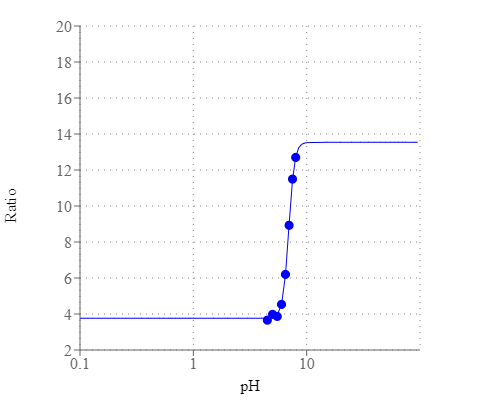
Intracellular pH (pHi) plays an important modulating role in many cellular events, including cell volume regulation, cellular metabolism, calcium regulation, receptor-mediated signal transduction, ion transport, endocytosis, and other cellular processes. Intracellular pH is generally 6.8 ~7.4 in the cytosol and 4.5~6.0 in the acidic organelles. Intracellular pH changes have significant physiological effects, e.g., the pH-dependent concentration of intracellular messengers such as Ca2+ and cAMP affects cellular signaling. Several recent reports showed the dysregulated pH is emerging as a hallmark of cancer cells. Spexyte™ Intracellular pH Calibration Buffer Kit provides a range of pH calibration buffers (pH 4.5~ 8.0) with nigericin, which modulate the intracellular pH with the external pH in the presence of 100"150 mM K+. When used in conjunction with pH indicators, such as BCFL, AM or BCECF, AM, Spexyte™ Intracellular pH Calibration Buffer Kit can create a standard curve which is used to determine the intracellular pH.

Platform


Fluorescence microscope

ExcitationTexas Red/FITC filter
EmissionTexas Red/FITC filter
Recommended plateBlack wall/clear bottom

Fluorescence microplate reader

Excitation440, 500 nm
Emission530 nm
Cutoff515 nm
Recommended plateBlack wall/clear bottom

Components


Example protocol


AT A GLANCE

Protocol Summary
  1. Stain cells with pH indicators (for example: BCFL,AM)
  2. Wash cells with HH Buffer
  3. Prepare Intracellular pH Calibration Buffer
  4. Add Intracellular pH Calibration Buffers to cells
  5. Incubate at 37 °C for 10 minutes
  6. Analyze the cells using the appropriate Ex/Em filter 
Important      HH Buffer and pH indicators are not provided in this kit.
Bring all the kit components to room temperature before starting the experiment.

CELL PREPARATION

For guidelines on cell sample preparation, please visit https://www.aatbio.com/resources/guides/cell-sample-preparation.html

PREPARATION OF STOCK SOLUTIONS

Unless otherwise noted, all unused stock solutions should be divided into single-use aliquots and stored at -20 °C after preparation. Avoid repeated freeze-thaw cycles.

Nigericin stock solution (10 mM)
Add 276 µL of DMSO (Component J) into the vial of Nigericin (Component I) to make a 10 mM Nigericin stock solution.

PREPARATION OF WORKING SOLUTION

Add 1 µL of 10 mM Nigericin stock solution into 1 mL standard pH buffer (Component A to Component H) to make Intracellular pH Calibration Buffer.

SAMPLE EXPERIMENTAL PROTOCOL

Stain cells with BCFL, AM
  1. Prepare 5 mM BCFL, AM (Cat#21190) in DMSO solution.
  2. Dilute to 5 µM in HH buffer + 0.02% PF127(Cat #20053).
  3. Remove growth medium from cells.
  4. Add 100 µL BCFL, AM staining solution.
  5. Incubate at 37 °C for 30 minutes.
  6. Remove BCFL, AM staining solution, and wash once with HH Buffer.
  7. Empty each well. 

Prepare Intracellular pH standard curve
  1. Add 100 µL Intracellular pH calibration buffer to cells.
  2. Incubate at 37 °C for at 5-10 minutes.
  3. Analyze the cells using the appropriate Ex/Em filters. For example: BCFL, AM: Ex= 440, 500nm, Em=530nm. 

Images


Citations


View all 9 citations: Citation Explorer
Empagliflozin suppressed cardiac fibrogenesis through sodium-hydrogen exchanger inhibition and modulation of the calcium homeostasis
Authors: Chung, Cheng-Chih and Lin, Yung-Kuo and Chen, Yao-Chang and Kao, Yu-Hsun and Yeh, Yung-Hsin and Trang, Nguyen Ngoc and Chen, Yi-Jen
Journal: Cardiovascular Diabetology (2023): 1--15
PHD2 is a regulator for glycolytic reprogramming in macrophages
Authors: Guentsch, Annemarie and Beneke, Angelika and Swain, Lija and Farhat, Katja and Nagarajan, Shunmugam and Wielockx, Ben and Raithatha, Kaamini and Dudek, Jan and Rehling, Peter and Zieseniss, Anke and others, undefined
Journal: Molecular and Cellular Biology (2016): MCB--00236
Design, calibration and application of broad-range optical nanosensors for determining intracellular pH
Authors: Sondergaard, R. V., Henriksen, J. R., Andresen, T. L.
Journal: Nat Protoc (2014): 2841-58
Manipulation of intracellular pH by electroporation: an alternative method for fast calibration of pH in living cells
Authors: Vecer, J., Holoubek, A., Herman, P.
Journal: Anal Biochem (2004): 348-50
Flow cytometric calibration of intracellular pH measurements in viable cells using mixtures of weak acids and bases
Authors: Chow, S., Hedley, D., Tannock, I.
Journal: Cytometry (1996): 360-7
Calibration methods and avoidance of errors in measurement of intracellular pH (pHcyt) using the indicator bis(carboxyethyl)-5(6)-carboxyfluorescein (BCECF) in human platelets
Authors: Valant, P. A., Haynes, D. H.
Journal: J Fluoresc (1992): 191-206
A novel method for absolute calibration of intracellular pH indicators
Authors: Eisner, D. A., Kenning, N. A., O'Neill, S. C., Pocock, G., Richards, C. D., Valdeolmillos, M.
Journal: Pflugers Arch (1989): 553-8
Intracellular calibration of a pH-sensitive dye in isolated, perfused salamander proximal tubules
Authors: Chaillet, J. R., Boron, W. F.
Journal: J Gen Physiol (1985): 765-94
Umbelliferone as an Intracellular pH-sensitive fluorescent indicator and blood-brain barrier probe: instrumentation, calibration, and analysis
Authors: Sundt, T. M., Jr., Anderson, R. E.
Journal: J Neurophysiol (1980): 60-75

References


View all 56 references: Citation Explorer
Monitoring phospholipid dynamics during phagocytosis: application of genetically-encoded fluorescent probes
Authors: Sarantis H, Grinstein S.
Journal: Methods Cell Biol (2012): 429
Phagocytosis and digestion of pH-sensitive fluorescent dye (Eos-FP) transfected E. coli in whole blood assays from patients with severe sepsis and septic shock
Authors: Schreiner L, Huber-Lang M, Weiss ME, Hohmann H, Schmolz M, Schneider EM.
Journal: J Cell Commun Signal (2011): 135
The application of fluorescent probes for the analysis of lipid dynamics during phagocytosis
Authors: Flannagan RS, Grinstein S.
Journal: Methods Mol Biol (2010): 121
Quantification of microsized fluorescent particles phagocytosis to a better knowledge of toxicity mechanisms
Authors: Leclerc L, Boudard D, Pourchez J, Forest V, Sabido O, Bin V, Palle S, Grosseau P, Bernache D, Cottier M.
Journal: Inhal Toxicol (2010): 1091
Analysis of macrophage phagocytosis: quantitative assays of phagosome formation and maturation using high-throughput fluorescence microscopy
Authors: Steinberg BE, Grinstein S.
Journal: Methods Mol Biol (2009): 45
Phagocytosis and postphagocytic reaction of cord blood and adult blood monocyte after infection with green fluorescent protein-labeled Escherichia coli and group B Streptococci
Authors: Gille C, Leiber A, Mundle I, Spring B, Abele H, Spellerberg B, Hartmann H, Poets Ch F, Orlikowsky TW.
Journal: Cytometry B Clin Cytom (2009): 271
A fluorescently tagged C-terminal fragment of p47phox detects NADPH oxidase dynamics during phagocytosis
Authors: Li XJ, Tian W, Stull ND, Grinstein S, Atkinson S, Dinauer MC.
Journal: Mol Biol Cell (2009): 1520
Analysis of phosphoinositide dynamics during phagocytosis using genetically encoded fluorescent biosensors
Authors: Cosio G, Grinstein S.
Journal: Methods Mol Biol (2008): 287
Development of a highly specific rhodamine-based fluorescence probe for hypochlorous acid and its application to real-time imaging of phagocytosis
Authors: Kenmoku S, Urano Y, Kojima H, Nagano T.
Journal: J Am Chem Soc (2007): 7313
The nonopsonic allogeneic cell phagocytosis of macrophages detected by flow cytometry and two photon fluorescence microscope
Authors: Liu GW, Ma HX, Wu Y, Zhao Y.
Journal: Transpl Immunol (2006): 220