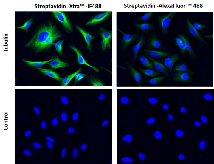
Streptavidin-Xtra™ IF488
Overview | ![]() ![]() |
Molecular weight 52000 | Correction Factor (260 nm) 0.21 | Correction Factor (280 nm) 0.11 | Extinction coefficient (cm -1 M -1) 750001 | Excitation (nm) 491 | Emission (nm) 516 | Quantum yield 0.91 |
Platform
Flow cytometer
Excitation | 488 nm laser |
Emission | 530/30 nm filter |
Instrument specification(s) | FITC channel |
Fluorescence microscope
Excitation | FITC filter set |
Emission | FITC filter set |
Recommended plate | Black wall/clear bottom |
Example protocol
PREPARATION OF STOCK SOLUTION
Streptavidin-Xtra™ stock solution (1 mg/mL):
Add 100 uL for Cat# 46000 and 1 mL for Cat# 46001 of ddH2O to make 1 mg/mL stock solution. Note: This reconstituted solution remains stable for 6 months without significant change when stored at 4 °C and kept from light. For longer storage, the reconstituted solution could be either divided into single-used aliquots, or added equal volume of glycerol (50% in final concentration) without aliquoting, and store the solution at -20°C (protected from light).
PREPARATION OF WORKING SOLUTION
Streptavidin-Xtra™ working solution:
For IF, the suggested staining concentration is at 1-5 ug/ml. For FACS, the suggested concentration is at 0.1-0.5 ug / 100 uL / million cells in staining buffer. Note: PBS + 0.1% BSA can be used as a staining buffer. Note: For the best performance of each application, the optimal concentration of this reagent needs to be carefully determined. Note: The suggested working dilution is provided as a guide only. It is recommended that the users titrate the product in their tests using proper positive and negative controls.
SAMPLE EXPERIMENTAL PROTOCOL
- Block and treat the samples with antibodies of interest as per the manufacturer's recommendations.
- Add biotin conjugated secondary antibody working solution in the samples at appropriate concentration and duration. Note: Please verify the compatibility and type of your biotin conjugated antibody with the primary antibody used in the experiment. For example, If the primary antibody is Mouse antibody then Goat Anti-Mouse antibody bound with biotin can be used for the assay.
- Incubate the cells with Streptavidin-Xtra™ working solution at room temperature for 30 minutes to 1 hour. Note: Optimal time for incubation needs to be determined carefully.
- Remove the working solution and resuspend the cells in your choice of buffer.
- Take the image using the fluorescence microscope or record the intensity using flow cytometer.
Calculators
Common stock solution preparation
0.1 mg | 0.5 mg | 1 mg | 5 mg | 10 mg | |
1 mM | 1.923 µL | 9.615 µL | 19.231 µL | 96.154 µL | 192.308 µL |
5 mM | 384.615 nL | 1.923 µL | 3.846 µL | 19.231 µL | 38.462 µL |
10 mM | 192.308 nL | 961.538 nL | 1.923 µL | 9.615 µL | 19.231 µL |
Molarity calculator
Mass (Calculate) | Molecular weight | Volume (Calculate) | Concentration (Calculate) | Moles | ||||
/ | = | x | = |
Spectrum
Open in Advanced Spectrum Viewer
Spectral properties
Correction Factor (260 nm) | 0.21 |
Correction Factor (280 nm) | 0.11 |
Extinction coefficient (cm -1 M -1) | 750001 |
Excitation (nm) | 491 |
Emission (nm) | 516 |
Quantum yield | 0.91 |
Images
Hela cells were fixed with 4% paraformaldehyde for 30 minutes, permeabilized with 0.02% Triton™ X-100 for 10 minutes, and blocked with 1% BSA for 1 hour. Fixed Hela cells were then stained with 1 µg/mL alpha Tubulin Mouse Monoclonal Antibody for 1 hour at room temperature, followed by GxM IgG-biotin (Cat# 16729) stain and then visualized with Streptavidin-Xtra™ iFluor 488 and Streptavidin-Alexa Fluor™ 488. Cell nuclei were stained with Hoechst 33342 (Blue, Cat#17535).
Citations
Authors: Wei, Y. P., Liu, X. P., Mao, C. J., Niu, H. L., Song, J. M., Jin, B. K.
Journal: Biosens Bioelectron (2018): 99-103
Authors: Yang, Z., Lan, Q., Li, J., Wu, J., Tang, Y., Hu, X.
Journal: Biosens Bioelectron (2017): 312-318
Authors: Hao, K., He, Y., Lu, H., Pu, S., Zhang, Y., Dong, H., Zhang, X.
Journal: Anal Chim Acta (2017): 114-120
Authors: Mishina, M., Minamihata, K., Moriyama, K., Nagamune, T.
Journal: Biomacromolecules (2017): 311
Authors: Guo, Q., Han, J. J., Shan, S., Liu, D. F., Wu, S. S., Xiong, Y. H., Lai, W. H.
Journal: Biosens Bioelectron (2016): 990-995
Authors: Chen, J., Liu, Y., Ji, X., He, Z.
Journal: Biosens Bioelectron (2016): 221-8
Authors: Lakshmipriya, T., Gopinath, S. C., Tang, T. H.
Journal: PLoS One (2016): e0151153
Authors: Mishina, M., Minamihata, K., Moriyama, K., Nagamune, T.
Journal: Biomacromolecules (2016): 1978-84
Authors: Mortberg, A., Meinke, S., Berg, P., Killie, M. K., Kjeldsen-Kragh, J., Jaras, K., Refsum, E., Hoglund, P., Wikman, A.
Journal: J Immunol Methods (2016): 9-15
Authors: Wu, D., Wang, Y., Zhang, Y., Ma, H., Pang, X., Hu, L., Du, B., Wei, Q.
Journal: Biosens Bioelectron (2016): 9-13