Amplite® Fluorimetric Beta-Hydroxybutyrate (Ketone Body) Assay Kit
| Price | |
| Catalog Number | |
| Unit Size | |
| Quantity |
| Telephone | 1-800-990-8053 |
| Fax | 1-800-609-2943 |
| sales@aatbio.com | |
| International | See distributors |
| Bulk request | Inquire |
| Custom size | Inquire |
| Shipping | Standard overnight for United States, inquire for international |
| H-phrase | H303, H313, H333 |
| Hazard symbol | XN |
| Intended use | Research Use Only (RUO) |
| R-phrase | R20, R21, R22 |
| UNSPSC | 12352200 |
| Overview |
Platform
Fluorescence microplate reader
| Excitation | 540 nm |
| Emission | 590 nm |
| Cutoff | 570 nm |
| Recommended plate | Solid black |
Components
Example protocol
AT A GLANCE
Protocol summary
- Prepare β-HB working solution (50 µL)
- Add β-HB standards or test samples (50 µL)
- Incubate at room temperature for 10 - 30 min
- Monitor fluorescence increase at Ex/Em = 540/590 nm
Important notes
To achieve the best results, it’s strongly recommended to use the black plates. Thaw one vial of each kit component at room temperature before starting the experiment.
PREPARATION OF STOCK SOLUTION
1. NAD stock solution (100X):
Add 100 µL of H2O into the vial of NAD (Component C) to make 100X NAD stock solution.
2. β-HB standard solution (100 mM):
Add 1 mL of H2O or 1X PBS buffer into the vial of β-HB standard (Component D) to make 100 mM β-HB standard solution.
PREPARATION OF STANDARD SOLUTION
For convenience, use the Serial Dilution Planner: https://www.aatbio.com/tools/serial-dilution/13831
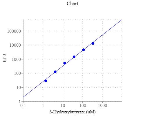
Add 10 µL of β-HB standard solution (100 mM) into 990 µL 1x PBS buffer to generate 1000 µM β-HB standard solution (HB7). Take the 1000 µM β-HB standard solution and perform 1:3 serial dilutions in 1x PBS to get serial dilutions of β-HB standard (HB6 - HB1). Note: Diluted β-HB standard solution is unstable and should be used within 4 hours.
PREPARATION OF WORKING SOLUTION
1. Add 5 mL of Assay Buffer (Component B) into one bottle of Enzyme Mix (Component A).
2. Add 50 µL NAD stock solution into the bottle of Component A+B, and mix well to make β-HB working solution (Component A+B+C). Note: This β-HB working solution is not stable, use it promptly and avoid direct exposure to light.
SAMPLE EXPERIMENTAL PROTOCOL
Table 1. Layout of β-HB standards and test samples in a clear bottom 96-well microplate. HB = β-HB standard (HB1 - HB7, 1 to 1000 µM); BL = blank control; TS = test sample.
| BL | BL | TS | TS |
| HB1 | HB1 | ... | ... |
| HB2 | HB2 | ... | ... |
| HB3 | HB3 | ||
| HB4 | HB4 | ||
| HB5 | HB5 | ||
| HB6 | HB6 | ||
| HB7 | HB7 |
Table 2. Reagent composition for each well.
| Well | Volume | Reagent |
| HB1-HB7 | 50 µL | Serial Dilution (1 to 1000 µM) |
| BL | 50 µL | 1X PBS Buffer |
| TS | 50 µL | Test Sample |
- Prepare β-HB standards (HB), blank control (BL) and test samples (TS) into a solid black 96-well microplate according to the layout provided in Tables 1 and 2. For a 384-well plate, use 25 µL of reagent per well instead of 50 µL.
- Add 50 µL of β-HB working solution to each well of β-HB standard, blank control, and test samples to make the total volume of 100 µL/well. For a 384-well plate, add 25 µL of β-HB working solution into each well instead, for a total volume of 50 µL/well.
- Incubate the reaction at room temperature for 10 - 30 minutes, protected from light.
- Monitor the fluorescence increase with a fluorescence plate reader at Excitation = 530 - 570 nm, Emission = 590 - 600 nm (optimal Ex/Em = 540/590 nm, cut off at 570 nm).
Images

Citations
Authors: Autterson, Gillian and Han, John Yeong Se and Philp, Nancy and Miller, Jason Matthew Lewis
Journal: Investigative Ophthalmology \& Visual Science (2023): 4466--4466
Authors: Zhang, Qitao and Presswalla, Feriel and Calton, Melissa and Charniga, Carol and Stern, Jeffrey and Temple, Sally and Vollrath, Douglas and Zacks, David N and Ali, Robin R and Thompson, Debra A and others,
Journal: Investigative ophthalmology \& visual science (2019): 3468--3479
Authors: Zhang, Qitao and Presswalla, Feriel and Feathers, Kecia and Cao, Xu and Hughes, Bret A and Zacks, David N and Thompson, Debra A and Miller, Jason ML
Journal: Experimental eye research (2018)
References
Authors: Wang W, Lan P.
Journal: J Biomater Sci Polym Ed (2014): 2094
Authors: Zharkova, II, Staroverova OV, Voinova VV, Andreeva NV, Shushckevich AM, Sklyanchuk ED, Kuzmicheva GM, Bespalova AE, Akulina EA, Shaitan KV, Olkhov AA.
Journal: Biomed Khim (2014): 553
Authors: Johansson J, Gronbladh A, Hallberg M.
Journal: Behav Brain Res (2014): 164
Authors: Wei T, Tian W, Liu F, Xie G.
Journal: Biochem Biophys Res Commun (2014): 666
Authors: Bonartsev AP, Yakovlev SG, Zharkova, II, Boskhomdzhiev AP, Bagrov DV, Myshkina VL, Makhina TK, Kharitonova EP, Samsonova OV, Feofanov AV, Voinova VV, Zernov AL, Efremov YM, Bonartseva GA, Shaitan KV, Kirpichnikov MP.
Journal: BMC Biochem (2013): 12
Authors: Cheng B, Lu H, Bai B, Chen J.
Journal: Neurochem Int (2013): 620
Authors: Lim J, Chong MS, Teo EY, Chen GQ, Chan JK, Teoh SH.
Journal: J Biomed Mater Res B Appl Biomater (2013): 752
Authors: Lomas AJ, Webb WR, Han J, Chen GQ, Sun X, Zhang Z, El Haj AJ, Forsyth NR.
Journal: Tissue Eng Part C Methods (2013): 577
Authors: Vogensen SB, Marek A, Bay T, Wellendorph P, Kehler J, Bundgaard C, Frolund B, Pedersen MH, Clausen RP.
Journal: J Med Chem (2013): 8201
Authors: Zheng J, Li D, Yuan L, Liu X, Chen H.
Journal: ACS Appl Mater Interfaces (2013): 5882
Application notes
Design of potent inhibitors of acetylcholinesterase using morin as the starting compound
Acetylcholinesterase Inhibitory Activity of Pigment Echinochrome A
Induction of Neurite Outgrowth in PC12 Cells
Induction of Neuritogenesis in PC12 Cells by a Pulsed Electromagnetic Field
FAQ
How should I reconstitute an NADPH standard?
Will Amplite® Fluorimetric NAD/NADH Ratio Assay Kit *Red Fluorescence* work with NADP/NADPH? Can this kit measure NADP+ and NADPH?
What is the concentration of calcium inside cells?
What assay kits measure NADP/NADPH from cell samples?