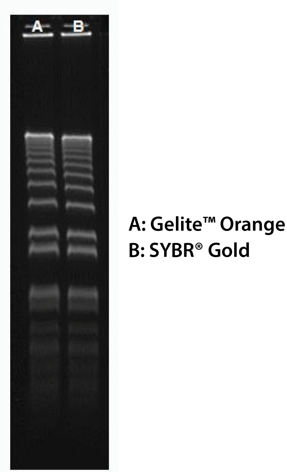
Gelite™ Orange Nucleic Acid Gel Staining Kit
Ordering information
Price | |
Catalog Number | |
Unit Size | |
Quantity |
Additional ordering information
Telephone | 1-800-990-8053 |
Fax | 1-800-609-2943 |
sales@aatbio.com | |
International | See distributors |
Bulk request | Inquire |
Custom size | Inquire |
Shipping | Standard overnight for United States, inquire for international |
Storage, safety and handling
H-phrase | H303, H313, H340 |
Hazard symbol | T |
Intended use | Research Use Only (RUO) |
R-phrase | R20, R21, R68 |
UNSPSC | 41116134 |
Alternative formats
Gelite™ Green Nucleic Acid Gel Staining Kit |
Related products
Overview | SDSProtocol |
See also: Cloning, DNA Fragmentation, DNA & RNA Markers and Ladders, Gel Electrophoresis, Loop-Mediated Isothermal Amplification (LAMP), Polymerase Chain Reaction (PCR), Reverse Transcription PCR (RT-PCR), RNA Purification & Analysis
Gelite™ Orange is an extremely sensitive nucleic acid gel stain for detecting DNA or RNA in gels using a standard 300 nm UV transilluminator and Polaroid 667 black-and-white print film. As with Helixyte™ Green stain, this remarkable sensitivity can be attributed to a combination of unique dye characteristics. Because the nucleic acid-bound Gelite™ Orange dye exhibits excitation maxima at both ~495 nm and ~300 nm (the emission maximum is ~537 nm), it is compatible with a wide variety of instrumentation, ranging from UV epi- and transilluminators and blue-light transilluminators, to mercury-arc lamp- and argon-ion laser-based gel scanners. Our Gelite™ Orange Nucleic Acid Gel Staining Gel Kit includes our Gelite™ Orange nucleic acid stain with an optimized and robust protocol. It provides a convenient solution for staining nucleic acid samples in gels.
Platform
Transilluminator
Excitation | 254 nm or 300 nm |
Emission | Long path green filter (ex. SYBR or GelStar) |
Components
Example protocol
PREPARATION OF WORKING SOLUTION
Add 1 μL of Gelite™ Orange (Component A) into 200 μL of 5X Gel Loading Buffer (Component B). Protect the working solution from light by covering it with foil or placing it in the dark.
SAMPLE EXPERIMENTAL PROTOCOL
- Prepare DNA samples as you desired.
- Add 4 µL of Gelite™ Orange working solution into 16 µL of DNA samples and mix well. Incubate at room temperature for 5 - 15 minutes prior to electrophoresis.
- Run gels based on your standard protocol.
- Image the gel with a 300 nm ultraviolet or 254 nm transilluminator, or a laser-based gel scanner using a long path green filter such as a SYBR® filter or GelStar® filter.
Images
References
View all 79 references: Citation Explorer
SYBR green real-time PCR for the detection of all enterovirus-A71 genogroups
Authors: Dubot-Peres A, Tan CY, de Chesse R, Sibounheuang B, Vongsouvath M, Phommasone K, Bessaud M, Gazin C, Thirion L, Phetsouvanh R, Newton PN, de Lamballerie X.
Journal: PLoS One (2014): e89963
Authors: Dubot-Peres A, Tan CY, de Chesse R, Sibounheuang B, Vongsouvath M, Phommasone K, Bessaud M, Gazin C, Thirion L, Phetsouvanh R, Newton PN, de Lamballerie X.
Journal: PLoS One (2014): e89963
Development of loop-mediated isothermal amplification and SYBR green real-time PCR methods for the detection of Citrus yellow mosaic badnavirus in citrus species
Authors: Anthony Johnson AM, Dasgupta I, Sai Gopal DV.
Journal: J Virol Methods (2014): 9
Authors: Anthony Johnson AM, Dasgupta I, Sai Gopal DV.
Journal: J Virol Methods (2014): 9
Development of both multiple PCR and real-time SYBR green PCR for the detection of Vibrio cholerae non-O1/O139 serogroups
Authors: Li F, Kan B, Wang D.
Journal: Zhonghua Liu Xing Bing Xue Za Zhi (2014): 66
Authors: Li F, Kan B, Wang D.
Journal: Zhonghua Liu Xing Bing Xue Za Zhi (2014): 66
Detection of Cardamom mosaic virus and Banana bract mosaic virus in cardamom using SYBR Green based reverse transcription-quantitative PCR
Authors: Siljo A, Bhat AI, Biju CN.
Journal: Virusdisease (2014): 137
Authors: Siljo A, Bhat AI, Biju CN.
Journal: Virusdisease (2014): 137
Comparison of SYBR Green and TaqMan methods in quantitative real-time polymerase chain reaction analysis of four adenosine receptor subtypes
Authors: Tajadini M, Panjehpour M, Javanmard SH.
Journal: Adv Biomed Res (2014): 85
Authors: Tajadini M, Panjehpour M, Javanmard SH.
Journal: Adv Biomed Res (2014): 85
Development of a High-resolution Melting Analysis Method Based on SYBR Green-I for rs7216389 Locus Genotyping in Asthmatic Child Patients
Authors: Vali Z, Raz A, Bokharaei H, Nabavi M, Bemanian MH, Yazdi MS, Djadid ND.
Journal: Avicenna J Med Biotechnol (2014): 72
Authors: Vali Z, Raz A, Bokharaei H, Nabavi M, Bemanian MH, Yazdi MS, Djadid ND.
Journal: Avicenna J Med Biotechnol (2014): 72
Primer evaluation and adaption for cost-efficient SYBR Green-based qPCR and its applicability for specific quantification of methanogens
Authors: Reitschuler C, Lins P, Illmer P.
Journal: World J Microbiol Biotechnol (2014): 293
Authors: Reitschuler C, Lins P, Illmer P.
Journal: World J Microbiol Biotechnol (2014): 293
Development and comparative evaluation of SYBR Green I-based one-step real-time RT-PCR assay for detection and quantification of West Nile virus in human patients
Authors: Kumar JS, Saxena D, Parida M.
Journal: Mol Cell Probes. (2014)
Authors: Kumar JS, Saxena D, Parida M.
Journal: Mol Cell Probes. (2014)
Multiplex SYBR(R) green-real time PCR (qPCR) assay for the detection and differentiation of Bartonella henselae and Bartonella clarridgeiae in cats
Authors: Staggemeier R, Pilger DA, Spilki FR, Cantarelli VV.
Journal: Rev Inst Med Trop Sao Paulo (2014): 93
Authors: Staggemeier R, Pilger DA, Spilki FR, Cantarelli VV.
Journal: Rev Inst Med Trop Sao Paulo (2014): 93
Development and evaluation of SYBR Green-I based quantitative PCR assays for herpes simplex virus type 1 whole transcriptome analysis
Authors: Garvey CE, McGowin CL, Foster TP.
Journal: J Virol Methods (2014): 101
Authors: Garvey CE, McGowin CL, Foster TP.
Journal: J Virol Methods (2014): 101
Application notes
A Novel Fluorescent Probe for Imaging and Detecting Hydroxyl Radical in Living Cells
Fluorescent Oligonucleotide Labeling Reagents
Monitoring of Mitochondrial Membrane Potential Changes in Live Cells Using JC-10
Selective Analysis of RNA in Live and Fixed Cells with StrandBrite RNA Green
Cell Loading Protocol For Fluorescent pH Indicator, BCECF-AM
Fluorescent Oligonucleotide Labeling Reagents
Monitoring of Mitochondrial Membrane Potential Changes in Live Cells Using JC-10
Selective Analysis of RNA in Live and Fixed Cells with StrandBrite RNA Green
Cell Loading Protocol For Fluorescent pH Indicator, BCECF-AM
FAQ
I ordered your phalloidin-amine (Cat #5302) so I can conjugate it to my oligo. Do you have a recommended protocol I can use?
What dye works best for staining and tracking lysosomes in live cells for several hours?
How can I lyse my cells without lysing the nuclear membrane?
Do you have any dual-fluorescence nucleic acid stains that interact with both DNA and RNA?
Do you have any fixable mitochondria staining assay kits?
What dye works best for staining and tracking lysosomes in live cells for several hours?
How can I lyse my cells without lysing the nuclear membrane?
Do you have any dual-fluorescence nucleic acid stains that interact with both DNA and RNA?
Do you have any fixable mitochondria staining assay kits?