Products
Services
Resources
Selection Guides
About
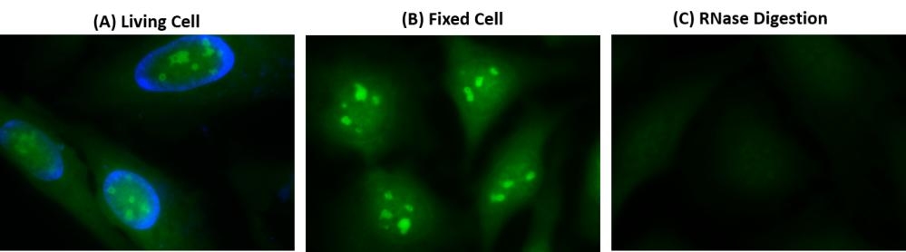
Cell Navigator® Live Cell RNA Imaging Kit
Green Fluorescence
Detecting and imaging RNA molecules in living cells is extremely important for a wide variety of molecular biology procedures including physical transportation, interpretation of genetic information, regulation of gene expression and some essential bio-catalytic roles. The major challenge to stain RNA in living cells is the interferences caused by DNA. In order to address the difficulty, a novel green fluorogenic dye was developed as a RNA-selective probe. AAT Bioquest's Cell Navigator® Live Cell RNA Imaging Kit includes StrandBrite™ RNA Green as it specifically binds RNA in cells. Compared to commercial SYTO™ RNA Select dye for RNA staining in vivo, StrandBrite™ RNA Green shows brighter signal and much better selectivity to RNA. In addition, this kit can stain RNA in both living cells and fixed cells.
| Catalog | Size | Price | Quantity |
|---|---|---|---|
| 22630 | 100 Tests | Price |

Spectral properties
| Excitation (nm) | 509 |
| Emission (nm) | 527 |
Usage and storage
| Intended use | Research Use Only (RUO) |
Instrument settings
| Fluorescence microscope | |
| Excitation | 490 nm |
| Emission | 520 nm |
| Recommended plate | Black wall/clear bottom |
| Instrument specification(s) | FITC filter set |
Contact us
| Telephone | |
| Fax | |
| sales@aatbio.com | |
| International | See distributors |
| Bulk request | Inquire |
| Custom size | Inquire |
| Technical Support | Contact us |
| Request quotation | Request |
| Purchase order | Send to sales@aatbio.com |
| Shipping | Standard overnight for United States, inquire for international |
Page updated on July 26, 2026