logo
AAT Bioquest

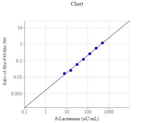
Amplite® Colorimetric Beta-Lactamase Activity Assay Kit

β-Lactamase dose response was measured with Amplite™ Colorimetric Beta-Lactamase Assay Kit (Cat#12551) on a 96-well clear bottom plate using a SpectraMax reader (Molecular Devices). 
Ordering information
Price
Catalog Number
Unit Size
Quantity
Add to cart
Additional ordering information
Telephone1-800-990-8053
Fax1-800-609-2943
Emailsales@aatbio.com
InternationalSee distributors
Bulk requestInquire
Custom sizeInquire
ShippingStandard overnight for United States, inquire for international
Request quotation
Storage, safety and handling
H-phraseH303, H313, H333
Hazard symbolXN
Intended useResearch Use Only (RUO)
R-phraseR20, R21, R22
UNSPSC12171501

OverviewpdfSDSpdfProtocol


β-Lactamases are a large family of enzymes capable of hydrolyzing β-lactams. β-Lactam ring is the common element in all beta-lactam antibiotics including penicillin derivatives, cephalosporins, monobactams, and carbapenems. Through hydrolysis, β-lactamase breaks the β-lactam ring open, thus deactivates the molecule's antibacterial properties. Bacteria from clinical and non-clinical settings are becoming increasingly resistant to β-lactam antibiotics by synthesizing β-lactamase. To overcome this resistance, β-lactam antibiotics are often given with β-lactamase inhibitors such as clavulanic acid. Therefore, detection of β-lactamase activity is of central importance to assess beta-lactam antibiotics as well as to prevent antibiotics resistance. AAT Bioquest's Colorimetric Beta-Lactamase Activity Assay Kit offers a sensitive colorimetric assay for measuring β-lactamase activity in biological samples. The β-lactamase activity is detected using Nitrocefin, which changes color from yellow to red upon hydrolysis by β-lactamase. The assay can be performed using an absorbance microplate reader by measuring the OD ratio at the wavelength of 490 nm to 380 nm.

Platform


Absorbance microplate reader

Absorbance490/380 nm
Recommended plateClear bottom

Components


Example protocol


AT A GLANCE

Protocol Summary
  1. Prepare β-Lactamase standards or test samples (50 µL)
  2. Add β-lactamase working solution (50 µL)
  3. Incubate at RT for 30 - 60 min
  4. Monitor absorbance increase at OD ratio of 490/380 nm 
Important      Thaw one vial of each kit component at room temperature before starting the experiment.

PREPARATION OF STOCK SOLUTIONS

Unless otherwise noted, all unused stock solutions should be divided into single-use aliquots and stored at -20 °C after preparation. Avoid repeated freeze-thaw cycles.

β-Lactamase standard stock solution (50 mU/mL)
Add 100 µL of ddH2O into the vial of β-Lactamase Standard (Component C) to make 50 mU/mL β-Lactamase standard solution.

PREPARATION OF STANDARD SOLUTION

For convenience, use the Serial Dilution Planner:
https://www.aatbio.com/tools/serial-dilution/12551


β-Lactamase standard
Add 10 µL of 50 mU/mL β-Lactamase standard solution into 990 µL 1x PBS buffer to generate 500 µU/mL β-Lactamase standard solution (SD7). Take 500 µU/mL β-Lactamase standard solution (SD7) and perform 1:2 serial dilutions in PBS to get serial diluted β-Lactamase standard (SD6 - SD1). Note: Diluted β-Lactamase standard solution is unstable, and should be used promptly.

PREPARATION OF WORKING SOLUTION

Add 50 µL Nitrocefin stock solution (Component A) into 5 mL of Assay Buffer (Component B ) and mix well to make β-Lactamase working solution. Note: This β-Lactamase working solution is enough for one 96-well plate. The β-Lactamase working solution is not stable, prepare fresh for each use.

SAMPLE EXPERIMENTAL PROTOCOL

Table 1.Layout of β-Lactamase standards and test samples in a 96-well clear bottom microplate. SD= β-Lactamase Standards (SD1 - SD7, 7.8 to 500 µU/mL), BL=Blank Control, TS=Test Samples.
BLBLTSTS
SD1SD1......
SD2SD2......
SD3SD3
SD4SD4
SD5SD5
SD6SD6
SD7SD7
Table 2. Reagent composition for each well.
WellVolumeReagent
SD1 - SD750 µLSerial Dilutions (7.8 to 500 µU/mL)
BL50 µL1x PBS Buffer
TS50 µLtest sample
  1. Prepare β-Lactamase standards (SD), blank controls (BL), and test samples (TS) according to the layout provided in Tables 1 and 2. For a 384-well plate, use 25 µL of reagent per well instead of 50 µL.
  2. Add 50 µL of β-Lactamase working solution to each well of β-Lactamase standard, blank control, and test samples to make the total β-Lactamase assay volume of 100 µL/well. For a 384-well plate, add 25 µL of β-Lactamase working solution into each well instead, for a total volume of 50 µL/well.
  3. Incubate the reaction at room temperature for 30 - 60 minutes, protected from light.
  4. Monitor the absorbance increase with an absorbance plate reader at OD ratio of 490/380 nm. 

Images


References


View all 14 references: Citation Explorer
A nitrocefin-based amperometric assay for the rapid quantification of extended-spectrum beta-lactamase-producing Escherichia coli in wastewaters
Authors: Chantemesse B, Betelli L, Solanas S, Vienney F, Bollache L, Hartmann A, Rochelet M.
Journal: Water Res (2017): 375
Amperometric detection of extended-spectrum beta-lactamase activity: application to the characterization of resistant E. coli strains
Authors: Rochelet M, Solanas S, Betelli L, Neuwirth C, Vienney F, Hartmann A.
Journal: Analyst (2015): 3551
Complete (1)H, (1)(5)N, and (1)(3)C resonance assignments of Bacillus cereus metallo-beta-lactamase and its complex with the inhibitor R-thiomandelic acid
Authors: Karsisiotis AI, Damblon C, Roberts GC.
Journal: Biomol NMR Assign (2014): 313
An altered zinc-binding site confers resistance to a covalent inactivator of New Delhi metallo-beta-lactamase-1 (NDM-1) discovered by high-throughput screening
Authors: Thomas PW, Spicer T, Cammarata M, Brodbelt JS, Hodder P, Fast W.
Journal: Bioorg Med Chem (2013): 3138
Evaluation of phenotypic tests for the detection of AmpC beta-lactamase in clinical isolates of Escherichia coli
Authors: H, undefined and a D, P and ey A, Asthana AK, Rawat A, H and a S, Thakuria B.
Journal: Indian J Pathol Microbiol (2013): 135
Horizontal Transfer of Antimicrobial Resistance by Extended-Spectrum beta Lactamase-Producing Enterobacteriaceae
Authors: Vaidya VK., undefined
Journal: J Lab Physicians (2011): 37
Prevalence of bla (CTX M) extended spectrum beta lactamase gene in enterobacteriaceae from critical care patients
Authors: Priyadharsini RI, Kavitha A, Rajan R, Mathavi S, Rajesh KR.
Journal: J Lab Physicians (2011): 80
Virtual screening of AmpC/beta-lactamase as target for antimicrobial resistance in Pseudomonas aeruginosa
Authors: Farmer R, Gautam B, Singh S, Yadav PK, Jain PA.
Journal: Bioinformation (2010): 290
Fine mapping of the sequence requirements for binding of beta-lactamase inhibitory protein (BLIP) to TEM-1 beta-lactamase using a genetic screen for BLIP function
Authors: Yuan J, Huang W, Chow DC, Palzkill T.
Journal: J Mol Biol (2009): 401
A sensitive coupled HPLC/electrospray mass spectrometry assay for SPM-1 metallo-beta-lactamase inhibitors
Authors: Sanchez PA, Toney JH, Thomas JD, Berger JM.
Journal: Assay Drug Dev Technol (2009): 170