logo
AAT Bioquest

Amplite® Colorimetric Enterokinase Activity Assay Kit

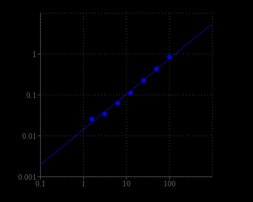
Enterokinase dose response was measured with Amplite® Colorimetric Enterokinase Activity Assay Kit (Cat #11410) on a 96-well clear bottom microplate using a SpectraMax microplate reader (Molecular Devices) with path check on mode.
Ordering information
Price
Catalog Number
Unit Size
Quantity
Add to cart
Additional ordering information
Telephone1-800-990-8053
Fax1-800-609-2943
Emailsales@aatbio.com
InternationalSee distributors
Bulk requestInquire
Custom sizeInquire
ShippingStandard overnight for United States, inquire for international
Request quotation
Storage, safety and handling
H-phraseH303, H313, H333
Hazard symbolXN
Intended useResearch Use Only (RUO)
R-phraseR20, R21, R22
UNSPSC12171501

OverviewpdfSDSpdfProtocol


Enterokinase (also called enteropeptidase) is a serine protease produced by cells in the duodenal wall and is a key enzyme in human and animal digestion system. Enterokinase converts trypsinogen into its active form trypsin, resulting in the subsequent activation of pancreatic digestive enzymes. The deficiency of enterokinase results in intestinal digestion impairment. The inhibition of enterokinase may have anti-tumor effects through suppressing proteases involved in carcinogenesis and metastasis. Therefore, highly selective and sensitive detection of enterokinase plays a key role in biochemical applications. Amplite® Colorimetric Enterokinase Activity Assay Kit offers a sensitive assay for quantifying enterokinase activity. After cleavage of enterokinase, the enterokinase substrate can be detected by EK Yellow™ in an absorbance microplate reader at 405 nm.
Platform


Absorbance microplate reader

Absorbance405 nm
Recommended plateClear bottom
Instrument specification(s)Path check on

Components


Example protocol


AT A GLANCE

Protocol summary

  1. Prepare test samples with diluted enterokinase standards (50 µL)
  2. Add equal volume of Enterokinase working solution (50 µL)
  3. Incubate at 37 °C for 30 - 60 minutes
  4. Monitor OD increase at 405 nm

Important notes
Thaw one vial of each kit component at room temperature before starting the experiment.

PREPARATION OF STOCK SOLUTION

Unless otherwise noted, all unused stock solutions should be divided into single-use aliquots and stored at -20 °C after preparation. Avoid repeated freeze-thaw cycles.

1. EK Yellow™ stock solution (100X):
Add 50 µL of DMSO (Component E) into EK Yellow™ (Component A) to make 100X stock solution.

2. Enterokinase Substrate stock solution (100X):
Add 50 µL of DMSO (Component E) into Enterokinase Substrate (Component B) to make 100X stock solution.

3. Enterokinase standard solution (10 ug/mL):
Add 50 uL of ddH2O + 0.1% BSA into Enterokinase Standard vial (Component D) to make 10 µg/mL Enterokinase stock solution.

PREPARATION OF STANDARD SOLUTION

Enterokinase standard

For convenience, use the Serial Dilution Planner: https://www.aatbio.com/tools/serial-dilution/11410

Add 10 µL of 10 µg/mL Enterokinase standard solution into 990 µL of Assay Buffer (Component C) to get 100 ng/mL enterokinase solution (EK7). Then perform 1:2 serial dilutions in assay buffer to get serially diluted enterokinase standards (EK6 - EK1). Note: The EK standards are for positive control only, and should not be relied on as a quantitation standard for enzyme activity.

PREPARATION OF WORKING SOLUTION

Add 50 µL of EK Yellow™ stock solution and 50 µL of Enterokinase Substrate stock solution into 5 mL of Assay Buffer (Component C); mix well to make Enterokinase (EK) working solution (Component A+B+C). Note: The assay mixture is enough for one 96-well plate. It is not stable, use promptly.

SAMPLE EXPERIMENTAL PROTOCOL

Table 1. Layout of enterokinase standards and test samples in a 96-well clear bottom microplate. EK = enterokinase standard (EK1 - EK7, 1.56 to 100 ng/mL); BL = blank control; TS = test sample.

BLBLTSTS
EK1EK1......
EK2EK2......
EK3EK3  
EK4EK4  
EK5EK5  
EK6EK6  
EK7EK7  

Table 2. Reagent composition for each well.

WellVolumeReagent
EK1 - EK750 µLserial dilution (1.56 to 100 ng/mL)
BL50 µLAssay Buffer (Component C)
TS50 µLsample
  1. Prepare enterokinase standards (EK), blank controls (BL), and test samples (TS) into a 96-well clear bottom microplate according to the layout provided in Table 1 and Table 2. For a 384-well plate, use 25 µL of reagent per well instead of 50 µL.

  2. Add 50 µL of EK working solution into each well of enterokinase standard, blank control, and test samples to make the total assay volume of 100 µL/well. For a 384-well plate, add 25 µL of EK working solution into each well instead, for a total volume of 50 µL/well.

  3. Incubate the reaction mixture at 37 °C for 30 - 60 minutes.

  4. Monitor the absorbance increase with an absorbance plate reader with path check on at OD of 405 nm.

Images


References


View all 23 references: Citation Explorer
Optimization of the fermentation and downstream processes for human enterokinase production in Pichia pastoris
Authors: Melicherova K, Krahulec J, Safranek M, Liskova V, Hopkova D, Szeliova D, Turna J.
Journal: Appl Microbiol Biotechnol. (2016)
Characterization of a gene family encoding SEA (sea-urchin sperm protein, enterokinase and agrin)-domain proteins with lectin-like and heme-binding properties from Schistosoma japonicum
Authors: Mbanefo EC, Kikuchi M, Huy NT, Shuaibu MN, Cherif MS, Yu C, Wakao M, Suda Y, Hirayama K.
Journal: PLoS Negl Trop Dis (2014): e2644
A strategy for fusion expression and preparation of functional glucagon-like peptide-1 (GLP-1) analogue by introducing an enterokinase cleavage site
Authors: Liu Y, Ren L, Ge L, Cui Q, Cao X, Hou Y, Bai F, Bai G.
Journal: Biotechnol Lett (2014): 1675
Do-it-yourself histidine-tagged bovine enterokinase: a handy member of the protein engineer's toolbox
Authors: Skala W, Goettig P, Br and stetter H., undefined
Journal: J Biotechnol (2013): 421
Fusion expression and purification of four disulfide-rich peptides reveals enterokinase secondary cleavage sites in animal toxins
Authors: Chen Z, Han S, Cao Z, Wu Y, Zhuo R, Li W.
Journal: Peptides (2013): 145
New strategy for specific activation of recombinant microbial pro-transglutaminase by introducing an enterokinase cleavage site
Authors: Wang K, Wang B, Yang HL, Pan L.
Journal: Biotechnol Lett (2013): 383
Immobilization of enterokinase on magnetic supports for the cleavage of fusion proteins
Authors: Santana SD, Pina AS, Roque AC.
Journal: J Biotechnol (2012): 378
Design and efficient production of bovine enterokinase light chain with higher specificity in E. coli
Authors: Chun H, Joo K, Lee J, Shin HC.
Journal: Biotechnol Lett (2011): 1227
The trail of my studies on glycoproteins from enterokinase to tumor markers
Authors: Yamashina I., undefined
Journal: Proc Jpn Acad Ser B Phys Biol Sci (2010): 578
Application of immobilized bovine enterokinase in repetitive fusion protein cleavage for the production of mucin 1
Authors: Kubitzki T, Minor D, Mackfeld U, Oldiges M, Noll T, Lutz S.
Journal: Biotechnol J (2009): 1610