logo
AAT Bioquest

Amplite® Colorimetric Glyceraldehyde-3-Phosphate Dehydrogenase (GAPDH) Assay Kit

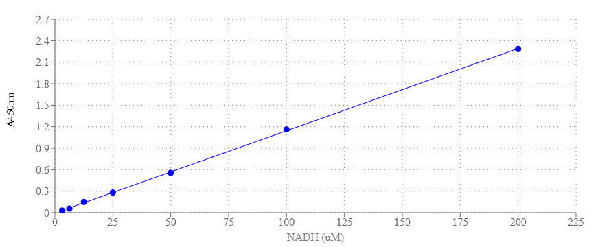
The Amplite® Colorimetric GAPDH Activity Assay Kit is a sensitive and simple tool for monitoring GAPDH activity based on a coupled enzymatic reaction catalyzed by GAPDH. This reaction results in the formation of a colorimetric product with an absorbance at 450nm, which is directly proportional to the enzymatic activity of GAPDH present in cell culture or tissue samples. One unit (U) is the amount of enzyme that catalyzes the reaction of 1 µmol of substrate per minute. GAPDH (Glyceraldehyde-3-Phosphate Dehydrogenase) is a multifunctional protein that serves both as a glycolytic enzyme and as a uracil DNA glycosylase. In glycolysis, it catalyzes the conversion of glyceraldehyde-3-phosphate to 1,3-bisphosphate glycerate. GAPDH is also involved in many cellular processes such as apoptosis, membrane trafficking, glucose and iron metabolism, and nuclear translocation. The expression of GAPDH in cells is constitutive, making it a housekeeping gene. Dysregulation of GAPDH activity has been associated with carcinogenesis, abnormal cell growth, and late-onset Alzheimer's disease. Quantification of GAPDH expression levels in different experimental conditions or disease states can provide insights into metabolic changes associated with cellular processes such as proliferation, differentiation, and response to stress.

Example protocol

AT A GLANCE

Important Note

Thaw all the kit components at room temperature before starting the experiment.

Protocol Summary
  1. Prepare the test samples, GAPDH Positive Control, and the serially diluted NADH standards (50 μL).

  2. Add the GAPDH working solution (50 μL).

  3. Incubate at room temperature for 10-30 minutes.

  4. Measure the absorbance at 450 nm.

PREPARATION OF STOCK SOLUTIONS

Unless otherwise noted, all unused stock solutions should be divided into single-use aliquots and stored at -20 °C after preparation. Avoid repeated freeze-thaw cycles

NADH Standard Stock Solution
  1. Add 200 µL of PBS buffer to the vial of NADH standard (Component D) to make a 2 mM (2 nmol/µL) NADH stock solution. Store the solution at -80°C. Avoid repeated freeze/thaw cycles.

GAPDH Positive Control Stock Solution
  1. To prepare a GAPDH stock solution, reconstitute the GAPDH Positive Control (Component E) by adding 40 µL of ddH2O. Mix well by pipetting and store at –20 °C.

    Note: Must be used within 2 months of reconstitution.

50X GAPDH Substrate Stock Solution
  1. To prepare a 50X GAPDH Substrate stock solution, add 100 µL of ddH2O to the vial containing the GAPDH Substrate (Component C). After mixing, store the solution at -20°C. Avoid repeated freezing and thawing.

PREPARATION OF STANDARD SOLUTIONS

For convenience, use the Serial Dilution Planner:
https://www.aatbio.com/tools/serial-dilution/11321

NADH Standard
Add 3 μL of 2 mM NADH standard solution to 297 μL of PBS + 0.1% BSA Buffer to prepare a 200 μM NADH solution (STD7). Then, take 150 μL of STD7 and perform 1:2 serial dilutions with PBS + 0.1% BSA Buffer to create a series of NADH standards from STD7 to STD1.

PREPARATION OF WORKING SOLUTION

GAPDH Working Solution
  1. Add 1 mL of the GAPDH Probe (Component B) to the bottle containing 4 mL of the GAPDH Assay Buffer (Component A), and mix well.

  2. Add 50 µL of the GAPDH Substrate stock solution to the same bottle and mix well.

    Note: This GAPDH working solution should be freshly prepared before each experiment and protected from light. A 5 mL solution is enough for 100 tests. Please prepare the necessary amount of GAPDH working solution based on this proportion.

SAMPLE EXPERIMENTAL PROTOCOL

GAPDH Positive Control
  1. Prepare one or more GAPDH positive control samples along with the test sample. The recommended concentration for the GAPDH positive control is 50 mU/mL in PBS + 0.1% BSA.

Table 1. Layout of GAPDH standards and test samples in a 96-well clear bottom microplate. (STD = NADH Standards (STD1-STD7, 3.125 to 200 uM), BL= Blank Control, TS = Test Samples.)

BL
BL
Positive Control
TS
STD 1
STD 1
...
...
STD 2
STD 2
...
...
STD 3
STD 3
STD 4
STD 4
STD 5
STD 5
STD 6
STD 6
STD 7
STD 7

Table 2. Reagent composition for each well.

Well
Volume
Reagent
STD 1 -STD 7
50 µL
NADH Serial Dilutions (3.125 to 200 µM)
BL
50 µL
PBS
GAPDH Positive Control
50 µL
GAPDH Positive Control
TS
50 µL
Test Sample
  1. Prepare the NADH standards (STD1-7), blank controls (BL), GAPDH Positive Control, and test samples (TS) according to the layout provided in Tables 1 and 2. When using a 384-well plate, use 25 µL of reagent per well instead of 50 µL.

  2. Add 50 µL of GAPDH Working Solution to each well containing the NADH standard, blank control, GAPDH Positive Control, and test samples. For a 384-well plate, add 25 µL of GAPDH Working Solution to each well instead.

  3. Incubate at room temperature for 10–30 minutes, protected from light.

  4. Monitor the absorbance intensity with an absorbance microplate reader at 450 nm.

References

View all 50 references: Citation Explorer
Triplet state spectroscopy reveals involvement of the buried tryptophan residue 310 in Glyceraldehyde-3-phosphate dehydrogenase (GAPD) in the interaction with acrylamide.
Authors: Mukherjee, Priyanka and Mukhopadhyay, Titas Kumar and Mukherjee, Manini and Roy, Pritam and Ghosh, Rina and Sardar, Pinki Saha and Ghosh, Sanjib
Journal: Spectrochimica acta. Part A, Molecular and biomolecular spectroscopy (2024): 123622
Structure-based functional analysis of a novel NADPH-producing glyceraldehyde-3-phosphate dehydrogenase from Corynebacterium glutamicum.
Authors: Son, Hyeoncheol Francis and Park, Woojin and Kim, Sangwoo and Kim, Il-Kwon and Kim, Kyung-Jin
Journal: International journal of biological macromolecules (2024): 128103
Structure of glyceraldehyde-3-phosphate dehydrogenase from Paracoccidioides lutzii in complex with an aldonic sugar acid.
Authors: Hernández-Prieto, Jonathan Heiler and Martini, Viviane Paula and Iulek, Jorge
Journal: Biochimie (2024): 20-33
Glyceraldehyde 3-Phosphate Dehydrogenase on the Surface of Candida albicans and Nakaseomyces glabratus Cells-A Moonlighting Protein That Binds Human Vitronectin and Plasminogen and Can Adsorb to Pathogenic Fungal Cells via Major Adhesins Als3 and Epa6.
Authors: Bednarek, Aneta and Satala, Dorota and Zawrotniak, Marcin and Nobbs, Angela H and Rapala-Kozik, Maria and Kozik, Andrzej
Journal: International journal of molecular sciences (2024)
Role of glyceraldehyde-3-phosphate dehydrogenase (GAPDH) in autophagy activation following subarachnoid hemorrhage.
Authors: Huo, Junfeng and Dong, Wei and Xu, Jiake and Ma, Lu and You, Chao
Journal: Experimental neurology (2024): 114577
Page updated on May 13, 2025

Ordering information

Price
Unit size
Catalog Number11321
Quantity
Add to cart

Additional ordering information

Telephone1-800-990-8053
Fax1-800-609-2943
Emailsales@aatbio.com
InternationalSee distributors
Bulk requestInquire
Custom sizeInquire
Technical SupportContact us
Purchase orderSend to sales@aatbio.com
ShippingStandard overnight for United States, inquire for international
Request quotation

Storage, safety and handling

H-phraseH303, H313, H333
Hazard symbolXN
Intended useResearch Use Only (RUO)
R-phraseR20, R21, R22
UNSPSC12352200

Platform

Absorbance microplate reader

Absorbance450 nm
Recommended plateClear bottom

Components

The NADH dose response was measured using the Amplite® Colorimetric Glyceraldehyde-3-Phosphate Dehydrogenase (GAPDH) Assay Kit on a 96-well clear bottom microplate. The assay was incubated for 10 minutes and measured at 450 nm using a ClarioStar microplate reader (BMG).