logo
AAT Bioquest

Amplite® Colorimetric NADP/NADPH Ratio Assay Kit

Amplite® Colorimetric NADP/NADPH Ratio Assay Kit is used to measure total NADP/NADPH amount in a white/clear 96-well microplate using a SpectraMax microplate reader (Molecular devices).
Amplite® Colorimetric NADP/NADPH Ratio Assay Kit is used to measure total NADP/NADPH amount and NADP/NADPH ratio in a white/clear 96-well microplate using a SpectraMax microplate reader (Molecular devices). Left: Total NADPH and NADP dose response; Right: NADP/NADPH ratio: Equal amount of NADP and NADPH mixture was treated with or without NADP extraction solution for 15 minutes, and then neutralized with extraction solution at room temperature. The signal was read at 460 nm. NADP/NADPH molar ratio is calculated based on the absorbance shown in the figure on the right.
Ordering information
Price
Catalog Number
Unit Size
Quantity
Add to cart
Additional ordering information
Telephone1-800-990-8053
Fax1-800-609-2943
Emailsales@aatbio.com
InternationalSee distributors
Bulk requestInquire
Custom sizeInquire
ShippingStandard overnight for United States, inquire for international
Request quotation
Storage, safety and handling
H-phraseH303, H313, H333
Hazard symbolXN
Intended useResearch Use Only (RUO)
R-phraseR20, R21, R22
UNSPSC12352200

OverviewpdfSDSpdfProtocol


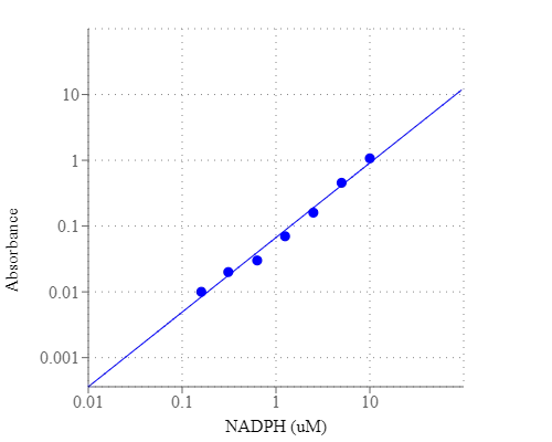
Nicotinamide adenine dinucleotide (NAD+) and nicotinamide adenine dinucleotide phosphate (NADP+) are two important cofactors found in cells. NADH is the reduced form of NAD+, and NAD+ is the oxidized form of NADH. It forms NADP with the addition of a phosphate group to the 2' position of the adenyl nucleotide through an ester linkage. NADP is used in anabolic biological reactions, such as fatty acid and nucleic acid synthesis, which require NADPH as a reducing agent. The traditional NAD/NADH and NADP/NADPH assays are done by monitoring of NADH or NADPH absorption at 340 nm. This method suffers low sensitivity and high interference since the assay is done in the UV range that requires expensive quartz microplate. Our Amplite® NADP/NADPH Ratio Assay Kit provides a convenient method for sensitive detection of NADP, NADPH and their ratio. The NADPH probe is a chromogenic sensor that has its maximum absorbance at ~460 nm upon NADH reduction. The absorbance increase at ~460 nm is directly proportional to the concentration of NADPH in the solution. The NADPH probe can recognize NADPH in an enzyme-free reaction, and the signal can be easily read by an absorbance microplate reader at ~460 nm. The Amplite® Colorimetric NADPH Assay Kit provides a sensitive assay to detect as little as 3 µM NADPH in a 100 µL assay volume. The assay can be performed in a convenient 96-well or 384-well microtiter-plate format.

Platform


Absorbance microplate reader

Absorbance460 nm
Recommended plateClear bottom

Components


Example protocol


AT A GLANCE

Protocol Summary
  1. Prepare 25 µL of NADPH standards and/or test samples
  2. Add 25 µL of NADP Extraction Solution
  3. Incubate at 37 °C for 15 minutes
  4. Add 25 µL of Neutralization Solution
  5. Add 75 µL of NADP/NADPH working solution
  6. Incubate at RT for 15 minutes to 2 hours
  7. Monitor Absorbance at 460 nm
Important Note
  1. It is highly recommended to incubate the cells with Lysis Buffer (Component G) at 37 °C and use the supernatant for the experiment.
  2. Thaw one of each kit component at room temperature before starting the experiment.

CELL PREPARATION

For guidelines on cell sample preparation, please visit https://www.aatbio.com/resources/guides/cell-sample-preparation.html

PREPARATION OF STOCK SOLUTIONS

Unless otherwise noted, all unused stock solutions should be divided into single-use aliquots and stored at -20 °C after preparation. Avoid repeated freeze-thaw cycles

NADPH standard solution (1 mM)

Add 200 µL of PBS buffer into the vial of NADPH standard (Component C) to have 1 mM (1 nmol/µL) NADPH stock solution.

PREPARATION OF STANDARD SOLUTIONS

For convenience, use the Serial Dilution Planner:
https://www.aatbio.com/tools/serial-dilution/15274

NADPH standard
Add 10 µL of 1 mM NADPH stock solution into 490 µL PBS buffer (pH 7.4) to generate 20 µM (20 pmols/µL) NADPH standard solution. Then take the 20 µM NADPH standard solution and perform 1:2 serial dilutions to get remaining serially diluted NADPH standards (NS7 - NS1). Note: Diluted NADPH standard solution is unstable, and should be used within 4 hours.

PREPARATION OF WORKING SOLUTION

  1. Add 8 mL of NADPH Probe buffer (Component B-II) to the bottle of NADP/NADPH Recycling Enzyme Mixture (Component A), and mix well.
  2. Add 2 mL NADPH Probe (Component B-I) into the same bottle (from Step 1) and mix well.

    Note        This NADP/NADPH working solution is enough for 125-200 assays. The working solution is not stable, use it promptly and avoid direct exposure to light.

SAMPLE EXPERIMENTAL PROTOCOL

TOTAL NADP+NADPH assay (avail. 400 assays/kit):

Table 1. Layout of NADPH standards and test samples in a white/clear bottom 96-well microplate. NS= NADPH Standards (NS1-NS7, 0.156 to 10 µM), BL=Blank Control, TS=Test Samples. 

BLBLTSTS
NS1NS1......
NS2NS2......
NS3NS3  
NS4NS4  
NS5NS5  
NS6NS6  
NS7NS7  

Table 2. Reagent composition for each well.
Note         High concentration of NADPH (e.g., >30 µM, final concentration) will cause saturated signal and make the calibration curve non-linear.

WellVolumeReagent
NS1 - NS750 µLSerial Dilutions (0.156 to 10 µM)
BL50 µLPBS
TS50 µLTest Sample
  1. Prepare NADPH standards (NS), blank controls (BL), and test samples (TS) according to the layout provided in Tables 1 and 2.

    Note         Prepare cells or tissue samples as desired. Lysis Buffer (Component G) can be used for lysing the cells for convenience.

    Note         It is highly recommended to incubate the cells with Lysis Buffer (Component G) at 37oC and use the supernatant for the experiment.
  2. Add 50 µL of NADP/NADPH working solution into each well of NADPH standard, blank control, and test samples to make the total NADP/NADPH assay volume of 100 µL/well.
  3. Incubate the reaction at room temperature for 15 minutes to 2 hours, protected from light. (We used 1 hour incubation time in data shown)
  4. Monitor the absorbance increase with an absorbance plate reader at 460 nm.
NADP/NADPH RATIO assay (avail. 250 assays/kit):

Table 3. Layout of NADPH standards and test samples in a white/clear 96-well microplate. NS= NADP/NADPH Standards (NS1 - NS7, 0.156 to 10 µM); BL=Blank Control; TS=Test Samples; TS (NADP) = Test Samples treated with NADP Extraction Solution (Component D) for 15 minutes, then neutralized by Neutralization Solution (Component E).

BLBLTSTSTS (NADP)TS (NADP)
NS1NS1......  
NS2NS2......  
NS3NS3    
NS4NS4    
NS5NS5    
NS6NS6    
NS7NS7    

Table 4. Reagent compositions for each well.
Note         High concentration of NADPH (e.g., >30 µM, final concentration) will cause saturated signal and make the calibration curve non-linear.

NADPH StandardBlank Control

Test Sample (NADP+NADPH)

Test Sample (NADP Extract)
Serial Dilutions: 25 µLPBS: 25 µLTest Sample: 25 µLTest Sample: 25 µL
Component F: 25 µLComponent F: 25 µLComponent F: 25 µLComponent D: 25 µL
Incubate at 37oC for 15 minutesIncubate at 37oC for 15 minutesIncubate at 37oC for 15 minutesIncubate at 37oC for 15 minutes
Component F: 25 µLComponent F: 25 µLComponent F: 25 µLComponent E: 25 µL
Total: 75 µLTotal: 75 µLTotal: 75 µLTotal: 75 µL
  1. Refer to Tables 3 & 4 for compositions of each well.  
    1. For NADP Extraction (NADP amount): Add 25 µL of NADP Extraction Solution (Component D) into the wells of NADP/NADPH containing test samples. Incubate at 37 °C for 10 to 15 minutes, then add 25 µL of Neutralization Solution (Component E) to neutralize the NADP extracts as described in Tables 3 & 4.
    2. For Total NADP and NADPH (Total amount): Add 25 µL of NADP/NADPH Control Solution (Component F) into the wells of NADPH standards and NADP/NADPH containing test samples. Incubate at 37o C for 10 to 15 minutes, and then add 25 µL of Extraction Control Solution (Component F) as described in Tables 3 and 4. 

      Note        Prepare cells or tissue samples as desired. Lysis Buffer (Component G) can be used for lysing the cells for convenience.
  2. Add 75 µL of NADP/NADPH working solution into each well of NADPH standard, blank control, and test samples (NADP/NADPH), and test sample (NADP Extract) to make the total assay volume of 150 µL/well.
  3. Incubate the reaction at room temperature for 15 minutes to 2 hours, protected from light.
  4. Monitor the absorbance increase with an absorbance plate reader at 460 nm.

Images


Citations


View all 67 citations: Citation Explorer
Detachment-activated FAK-STAT3-NNMT inhibits anoikis by enhancing fatty acid oxidation in breast cancer
Authors: Tong, Qingchao and Ma, Yilei and Gao, Yuzhen and Zhou, Lin and Fang, Qiang and Fang, Sining and Zhao, Ru and Zeng, Jin and Li, Guoli and Xie, Xinyou and others,
Journal: (2024)
Systematic Engineering to Enhance 8-Hydroxygeraniol Production in Yeast
Authors: Wang, Herong and Jiang, Guozhen and Liang, Nan and Dong, Tianyu and Shan, Mengying and Yao, Mingdong and Wang, Ying and Xiao, Wenhai and Yuan, Yingjin
Journal: Journal of Agricultural and Food Chemistry (2023)
Tumor-Associated Neutrophils Dampen Adaptive Immunity and Promote Cutaneous Squamous Cell Carcinoma Development
Authors: Khou, Sokchea and Popa, Alexandra and Luci, Carmelo and Bihl, Franck and Meghraoui-Kheddar, Aida and Bourdely, Pierre and Salavagione, Emie and Cosson, Estelle and Rubod, Alain and Cazareth, Julie and others,
Journal: Cancers (2020): 1860
Glsnf1-mediated metabolic rearrangement participates in coping with heat stress and influencing secondary metabolism in Ganoderma lucidum
Authors: Hu, Yanru and Xu, Wenzhao and Hu, Shishan and Lian, Lingdan and Zhu, Jing and Ren, Ang and Shi, Liang and Zhao, Ming Wen
Journal: Free Radical Biology and Medicine (2019)
Transcription factor YY1 promotes cell proliferation by directly activating the pentose phosphate pathway
Authors: Wu, Shourong and Wang, Huimin and Li, Yanjun and Xie, Yudan and Huang, Can and Zhao, Hezhao and Miyagishi, Makoto and Kasim, Vivi
Journal: Cancer research (2018): 4549--4562
Enhanced 1, 3-propanediol production in Klebsiella pneumoniae by a combined strategy of strengthening the TCA cycle and weakening the glucose effect
Authors: Lu, Xinyao and Ren, Shunli and Lu, Jingzheng and Zong, Hong and Song, Jian and Zhuge, Bin
Journal: Journal of applied microbiology (2018)
Enhancement of erythritol production by Trichosporonoides oedocephalis ATCC 16958 through regulating key enzyme activity and the NADPH/NADP ratio with metal ion supplementation
Authors: Li, Liangzhi and Kang, Pei and Ju, Xin and Chen, Jiajia and Zou, Huibin and Hu, Cuiying and Yan, Lishi
Journal: Preparative Biochemistry and Biotechnology (2018): 257--263
Resveratrol attenuates excessive ethanol exposure induced insulin resistance in rats via improving NAD+/NADH ratio
Authors: Luo, Gang and Huang, Bingqing and Qiu, Xiang and Xiao, Lin and Wang, Ning and Gao, Qin and Yang, Wei and Hao, Liping
Journal: Molecular Nutrition & Food Research (2017)