logo
AAT Bioquest

Amplite® Colorimetric Urea Quantitation Kit *Blue Color*

Urea dose response in a 96-well clear bottom plate using a SpectraMax microplate reader (Molecular Devices) measured with Amplite® Colorimetric Urea Assay Kit.
Ordering information
Price
Catalog Number
Unit Size
Quantity
Add to cart
Additional ordering information
Telephone1-800-990-8053
Fax1-800-609-2943
Emailsales@aatbio.com
InternationalSee distributors
Bulk requestInquire
Custom sizeInquire
ShippingStandard overnight for United States, inquire for international
Request quotation
Storage, safety and handling
H-phraseH303, H313, H333
Hazard symbolXN
Intended useResearch Use Only (RUO)
R-phraseR20, R21, R22
UNSPSC12171501

OverviewpdfSDSpdfProtocol


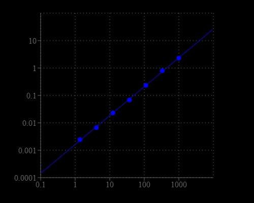
Urea is the final degradation product of protein and amino acid metabolism in animals. It is produced in liver, secreted by kidney and excreted through urine. The determination of urea is very useful test in clinical laboratory to monitor health status. The Blood Urea Nitrogen (BUN) test is a measure of the amount of nitrogen in the blood in the form of urea and is primarily used, along with the creatinine test to evaluate kidney function, helping diagnose kidney diseases. Our Amplite® Colorimetric Urea Assay Kit provides a simple and sensitive colorimetric method for the quantitation of urea concentration in biological samples such as serum, plasma and urine, etc. The assay is based on an enzyme-coupled reaction of urea in the assay buffer, and finally produces a blue colored product. The intensity of color produced is proportional to the concentration of urea in the sample, which can be measured colorimetrically at 660-670 nm. This Amplite® Colorimetric Urea Assay Kit provides a simple assay to detect as little as 10 µM urea in a 150 µL assay volume. The assay can be performed in a convenient 96-well or 384-well microtiter-plate format and easily adapted to automation without a separation step.

Platform


Absorbance microplate reader

Absorbance665 nm
Recommended plateClear bottom

Components


Example protocol


AT A GLANCE

Protocol summary

  1. Prepare urea standards or test samples (50 µL)
  2. Add urea working solution (50 µL)
  3. Incubate at room temperature or 37°C for 30 - 60 min
  4. Add Assay Buffer II
  5. Read Absorbance at 665 nm

Important notes
Thaw all the kit components to room temperature before starting the experiment.

PREPARATION OF STOCK SOLUTION

Unless otherwise noted, all unused stock solutions should be divided into single-use aliquots and stored at -20 °C after preparation. Avoid repeated freeze-thaw cycles.

1. Assay Enzyme Mix stock solution (100X):
Add 100 µL of ddH2O into the vial of Assay Enzyme Mix (Component A) to make 100X Assay Enzyme Mix stock solution.

PREPARATION OF STANDARD SOLUTION

Urea standard

For convenience, use the Serial Dilution Planner: https://www.aatbio.com/tools/serial-dilution/10058

Add 1 μL of 1.0 M Urea Standard (Component D) to 999 µL of DPBS to generate 1.0 mM standard urea solution (US7). Take 1.0 mM urea standard solution to perform 1:3 serial dilutions to get remaining urea standard solutions (US6 - US1).

PREPARATION OF WORKING SOLUTION

Add 50 μL of reconstituted Assay Enzyme Mix stock solution (100X) into 5 mL Assay Buffer I (Component B) to make urea working solution. Note: The urea working solution should be used promptly and kept from light. The assay sensitivity will be decreased with longer storage time. Fresh urea working solution is recommended for the best result.

SAMPLE EXPERIMENTAL PROTOCOL

Table 1. Layout of urea standards and test samples in a clear bottom 96-well microplate. US= Urea Standards (US1 - US7, 1 to 1000 µM), BL=Blank Control, TS=Test Samples. 

BLBLTSTS
US1US1......
US2US2......
US3US3  
US4US4  
US5US5  
US6US6  
US7US7  

Table 2. Reagent composition for each well.

WellVolumeReagent
US1 - US750 µLSerial Dilutions (1 to 1000 µM)
BL50 µLDPBS
TS50 µLtest sample
  1. Prepare urea standards (US), blank controls (BL), and test samples (TS) according to the layout provided in Tables 1 and 2. For a 384-well plate, use 25 µL of reagent per well instead of 50 µL.

  2. Add 50 µL of urea working solution to each well of urea standard, blank control, and test samples to make the total urea assay volume of 100 µL/well. For a 384-well plate, add 25 µL of urea working solution into each well instead, for a total volume of 50 µL/well.

  3. Incubate the reaction for 30 - 60 minutes at room temperature or 37°C, protected from light.

  4. Add 50 µL of Assay Buffer II (Component C) to each well so that the total assay volume is 150 µL/well. For a 384-well plate, add 25 µL Assay Buffer II (Component C) to each well, for a total assay volume of 75 µL/well.

  5. Incubate at room temperature for 10 - 15 minutes, and monitor the absorbance increase at 660 - 670 nm using an absorbance microplate reader. Note: The color turns to yellow after Assay Buffer II (Component C) is added, and the wells with urea standard or samples will show blue-green color after incubation. The intensity of the color will reach the maximum in 15 - 30 minutes, and is proportional to the concentration of urea. Note: The final color is stable for ~1 hour in room temperature and the color intensity will decrease with time.

Images


References


View all 60 references: Citation Explorer
A novel and sensitive resonance scattering assay for detection of urea in serum coupled urease catalytic reaction and NH4+ associated particle reaction
Authors: Liang A, Qin H, Zhou L, Zhang Y, Ouyang H, Wang P, Jiang Z.
Journal: Bioprocess Biosyst Eng (2011): 639
Dimethyl formamide-free, urea-NaCl fluorescence in situ hybridization assay for Staphylococcus aureus
Authors: Lawson TS, Connally RE, Vemulpad S, Piper JA.
Journal: Lett Appl Microbiol. (2011)
Evaluation of diuron (3-[3,4-dichlorophenyl]-1,1-dimethyl urea) in a two-stage mouse skin carcinogenesis assay
Authors: Ferrucio B, Franchi CA, Boldrin NF, de Oliveira ML, de Camargo JL.
Journal: Toxicol Pathol (2010): 756
Sample prep for proteomics of breast cancer: proteomics and gene ontology reveal dramatic differences in protein solubilization preferences of radioimmunoprecipitation assay and urea lysis buffers
Authors: Ngoka LC., undefined
Journal: Proteome Sci (2008): 30
Improvement of the decision efficiency of the accuracy profile by means of a desirability function for analytical methods validation. Application to a diacetyl-monoxime colorimetric assay used for the determination of urea in transdermal iontophoretic extr
Authors: Rozet E, Wascotte V, Lecouturier N, Preat V, Dewe W, Boulanger B, Hubert P.
Journal: Anal Chim Acta (2007): 239
Immobilization of urease from pigeonpea (Cajanus cajan) on agar tablets and its application in urea assay
Authors: Mulagalapalli S, Kumar S, Kalathur RC, Kayastha AM.
Journal: Appl Biochem Biotechnol (2007): 291
New enzymatic assay for serum urea nitrogen using urea amidolyase
Authors: Kimura S, Iyama S, Yamaguchi Y, Kanakura Y.
Journal: J Clin Lab Anal (2003): 52
Utilization of lacrimal urea assay in the monitoring of hemodialysis: conditions, limitations and lacrimal arginase characterization
Authors: Farkas A, Vamos R, Bajor T, Mullner N, Lazar A, Hraba A.
Journal: Exp Eye Res (2003): 183
Enzymatic assay for determination of bicarbonate ion in plasma using urea amidolyase
Authors: Kimura S, Yamanishi H, Iyama S, Yamaguchi Y, Kanakura Y.
Journal: Clin Chim Acta (2003): 179
Stool antigen assay (HpSA) is less reliable than urea breath test for post-treatment diagnosis of Helicobacter pylori infection
Authors: Bilardi C, Biagini R, Dulbecco P, Iiritano E, Gambaro C, Mele MR, Borro P, Tessieri L, Zentilin P, Mansi C, Vigneri S, Savarino V.
Journal: Aliment Pharmacol Ther (2002): 1733