logo
AAT Bioquest

Amplite® Colorimetric Xanthine Assay Kit

Xanthine dose response was measured with Amplite® Colorimetric Xanthine Assay Kit in a 96-well clear bottom plate using a SpectraMax reader (Molecular Devices).
Ordering information
Price
Catalog Number
Unit Size
Quantity
Add to cart
Additional ordering information
Telephone1-800-990-8053
Fax1-800-609-2943
Emailsales@aatbio.com
InternationalSee distributors
Bulk requestInquire
Custom sizeInquire
ShippingStandard overnight for United States, inquire for international
Request quotation
Storage, safety and handling
H-phraseH303, H313, H333
Hazard symbolXN
Intended useResearch Use Only (RUO)
R-phraseR20, R21, R22
UNSPSC12352200

OverviewpdfSDSpdfProtocol


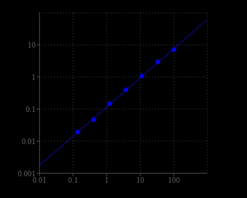
Xanthine is a purine base found in most human body tissues and fluids. A number of stimulants are derived from xanthine, including caffeine, aminophylline, IBMX, paraxanthine, pentoxifylline, theobromine, and theophylline, which can stimulate heart rate, force of contraction, cardiac arrhythmias at high concentrations. Therefore, detection of Xanthine alteration in biological samples is important for disease diagnosis and therapy monitoring. Amplite® Colorimetric Xanthine Assay Kit provides a quick and ultrasensitive method for the measurement of xanthine. It can be performed in a convenient 96-well or 384-well microtiter-plate format. Xanthine is oxidized to uric acid in the presence of xanthine oxidase to release hydrogen peroxide, which can be specifically measured with Amplite® Red by an absorbance microplate reader at 576 nm. With Amplite® Colorimetric Xanthine Assay Kit, as low as 1.2 µM xanthine was detected in a 100 µL reaction volume.

Platform


Absorbance microplate reader

Absorbance570/610 nm
Recommended plateClear bottom

Components


Example protocol


AT A GLANCE

Protocol Summary
  1. Prepare xanthine standards and test samples (50 µL)
  2. Add xanthine working solution (50 µL)
  3. Incubate at room temperature for 30 - 60 min
  4. Read absorbance increase at OD ratio of 570/610 nm 
Important      Thaw all the kit components at room temperature before starting the experiment.

PREPARATION OF STOCK SOLUTIONS

Unless otherwise noted, all unused stock solutions should be divided into single-use aliquots and stored at -20 °C after preparation. Avoid repeated freeze-thaw cycles.

1. Amplite™ Red Substrate stock solution (250X)
Add 40 µL of DMSO (Component F) into the vial of Amplite™ Red Substrate (Component A).
Note     Amplite™ Red Substrate is unstable in the presence of thiols such as dithiothreitol (DTT) and 2-mercaptoethanol. The final concentration of DTT or 2-mercaptoethanol in the reaction should be no higher than 10 µM. The assay should be performed at pH 7 – 8 (pH 7.4 is recommended) as Amplite™ Red is unstable pH >8.5.


2. HRP stock solution (500X)
Add 100 µL of Assay Buffer (Component B) into the vial of Horseradish Peroxidase (Component C).

3. Xanthine Oxidase (XO) stock solution (100X)
Add 100 µL of assay buffer (Component B) into the vial of Xanthine Oxidase Standard (Component E) to make Xanthine Oxidase (XO) stock solution (100X).

4. Xanthine standard solution (100 µM)
Add 5 µL of Xanthine Standard (Component D) into 995 µL of Assay Buffer (Component B) to get 100 µM Xanthine standard solution.

PREPARATION OF STANDARD SOLUTION

For convenience, use the Serial Dilution Planner:
https://www.aatbio.com/tools/serial-dilution/13842


Xanthine standard
Perform 1:3 serial dilution with 100 µM Xanthine standard solution (X7) to obtain Xanthine standards (X6 - X1).

PREPARATION OF WORKING SOLUTION

Add 20 μL of Amplite™ Red Substrate stock solution (250X), 10 μL of HRP stock solution (500X), and 50 μL of Xanthine Oxidase stock solution (100X) into 5 mL of Assay Buffer to make a total volume of 5.08 mL. Protect from light.

SAMPLE EXPERIMENTAL PROTOCOL

Table 1. Layout of Xanthine standards and test samples in a clear bottom 96-well microplate (50 µL/well). X= xanthine standards (X1 - X7, 0.1 to 100 µM), BL=blank control, TS=test samples.
BLBLTSTS
X1X1......
X2X2......
X3X3
X4X4
X5X5
X6X6
X7X7
Table 2. Reagent composition for each well.
WellVolumeReagent
X1 - X750 µLSerial Dilutions (0.1 to 100 µM)
BL50 µLAssay Buffer (Component B)
TS50 µLtest sample
  1. Prepare xanthine standards (X), blank controls (BL), and test samples (TS) according to the layout provided in Tables 1 and 2. For a 384-well plate, use 25 µL of reagent per well instead of 50 µL.
  2. Add 50 µL of working solution to each well of xanthine standard, blank control, and test samples to make the total xanthine assay volume of 100 µL/well. For a 384-well plate, add 25 µL of working solution into each well instead, for a total volume of 50 µL/well.
  3. Incubate the reaction for 30 to 60 minutes at room temperature, protected from light.
  4. Monitor signal intensity with an absorbance plate reader at OD ratio of 570/610 nm. 

Images


References


View all 60 references: Citation Explorer
Concentration-dependent effect of sodium hypochlorite on stem cells of apical papilla survival and differentiation
Authors: Martin DE, De Almeida JF, Henry MA, Khaing ZZ, Schmidt CE, Teixeira FB, Diogenes A.
Journal: J Endod (2014): 51
Effect of hypochlorite oxidation on cholinesterase-inhibition assay of acetonitrile extracts from fruits and vegetables for monitoring traces of organophosphate pesticides
Authors: Kitamura K, Maruyama K, Hamano S, Kishi T, Kawakami T, Takahashi Y, Onodera S.
Journal: J Toxicol Sci (2014): 71
Green synthesis of carbon dots with down- and up-conversion fluorescent properties for sensitive detection of hypochlorite with a dual-readout assay
Authors: Yin B, Deng J, Peng X, Long Q, Zhao J, Lu Q, Chen Q, Li H, Tang H, Zhang Y, Yao S.
Journal: Analyst (2013): 6551
Comparative antimicrobial activities of aerosolized sodium hypochlorite, chlorine dioxide, and electrochemically activated solutions evaluated using a novel standardized assay
Authors: Thorn RM, Robinson GM, Reynolds DM.
Journal: Antimicrob Agents Chemother (2013): 2216
Analysis of the germination kinetics of individual Bacillus subtilis spores treated with hydrogen peroxide or sodium hypochlorite
Authors: Setlow B, Yu J, Li YQ, Setlow P.
Journal: Lett Appl Microbiol (2013): 259
A simple yet effective chromogenic reagent for the rapid estimation of bromate and hypochlorite in drinking water
Authors: Zhang J, Yang X.
Journal: Analyst (2013): 434
Effect of hypochlorite-based disinfectants on inactivation of murine norovirus and attempt to eliminate or prevent infection in mice by addition to drinking water
Authors: Takimoto K, Taharaguchi M, Sakai K, Takagi H, Tohya Y, Yamada YK.
Journal: Exp Anim (2013): 237
Use of pyrogallol red and pyranine as probes to evaluate antioxidant capacities towards hypochlorite
Authors: Perez-Cruz F, Cortes C, Atala E, Bohle P, Valenzuela F, Olea-Azar C, Speisky H, Aspee A, Lissi E, Lopez-Alarcon C, Bridi R.
Journal: Molecules (2013): 1638
Enhancement of anti-cholinesterase activity of aqueous samples by hypochlorite oxidation for monitoring traces of organophosphorus pesticides in water
Authors: Kanno A, Kawakami T, Takahashi Y, Onodera S.
Journal: J Toxicol Sci (2012): 389
Colorimetric determination of hypochlorite with unmodified gold nanoparticles through the oxidation of a stabilizer thiol compound
Authors: Zhang J, Wang X, Yang X.
Journal: Analyst (2012): 2806