logo
AAT Bioquest

Amplite® Colorimetric Xanthine Oxidase Assay Kit

Xanthine oxidase dose response was measured with Amplite® Colorimetric Xanthine Oxidase Assay Kit in a white or black wall/clear bottom 96-well microplate using a SpectraMax microplate reader (Molecular Devices). As low as 0.12 mU/mL xanthine oxidase was detected with 30 minutes incubation time (n=3).
Ordering information
Price
Catalog Number
Unit Size
Quantity
Add to cart
Additional ordering information
Telephone1-800-990-8053
Fax1-800-609-2943
Emailsales@aatbio.com
InternationalSee distributors
Bulk requestInquire
Custom sizeInquire
ShippingStandard overnight for United States, inquire for international
Request quotation
Spectral properties
Excitation (nm)571
Emission (nm)584
Storage, safety and handling
H-phraseH303, H313, H333
Hazard symbolXN
Intended useResearch Use Only (RUO)
R-phraseR20, R21, R22
UNSPSC12171501

OverviewpdfSDSpdfProtocol


Excitation (nm)
571
Emission (nm)
584
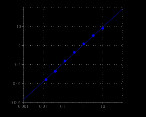
Xanthine oxidase (XO)is an enzyme that catalyzes the oxidation of hypoxanthine to xanthine and can further catalyze the oxidation of xanthine to uric acid. It plays an important role in the catabolism of purines. Xanthine oxidase is normally found in liver and jejunum. During severe liver damage, xanthine oxidase is released into blood, so a blood assay for XO is a way to determine if liver damage has happened. Xanthinuria is a rare genetic disorder where the lack of xanthine oxidase leads to high concentration of xanthine in blood and can cause health problems such as renal failure. The Amplite® Colorimetric Xanthine Oxidase Assay Kit provides a quick and ultrasensitive method for the measurement of xanthine oxidase activities. It can be performed in a convenient 96-well or 384-well microtiter-plate format and easily adapted to automation without a separation step. In the assay, xanthine oxidase catalyzes the oxidation of purine bases, hypoxanthine or xanthine to uric acid and superoxide , which spontaneously degrades to hydrogen peroxide (H2O2). The kit uses our Amplite® Red substrate which enables a dual recordable mode. The color signal can be easily read at ~570 nm with an absorbance microplate reader. With the Amplite® Colorimetric Xanthine Oxidase Assay Kit, we have detected as little as 0.3 mU/mL xanthine oxidase in a 100 µL reaction volume.

Platform


Absorbance microplate reader

Absorbance570/610 nm
Recommended plateClear bottom

Components


Example protocol


AT A GLANCE

Protocol Summary
  1. Prepare and add XO standards and/or test samples (50 µL)
  2. Prepare and add XO Assay working solution (50 µL)
  3. Incubate at room temperature for 30-60 minutes
  4. Read absorbance increase at OD ratio of 570/610 nm 
Important      Thaw all the kit components at room temperature before starting the experiment.

PREPARATION OF STOCK SOLUTIONS

Unless otherwise noted, all unused stock solutions should be divided into single-use aliquots and stored at -20 °C after preparation. Avoid repeated freeze-thaw cycles.

1. Amplite™ Red stock solution (250X)
Add 40 µL of DMSO (Component F) into the vial of Amplite™ Red substrate (Component A). The stock solution should be used promptly.
Note      The Amplite™ Red substrate is unstable in the presence of thiols such as dithiothreitol (DTT) and 2-mercaptoethanol. The final concentration of DTT or 2-mercaptoethanol in the reaction should be no higher than 10 µM. The Amplite™ Red substrate is also unstable at high pH (> 8.5). Therefore, the reaction should be performed at pH 7–8. The provided Assay Buffer (pH 7.4) is recommended.


2. HRP stock solution (500X)
Add 100 µL of Assay Buffer (Component B) into the vial of HRP (Component C).
Note      The unused HRP stock solution (500X) should be divided into single use aliquots and stored them at -20 oC.


3. Xanthine Oxidase (XO) stock solution
Add 200 µL of Assay Buffer (Component B) into the vial of Xanthine Oxidase Standard (Component E).
Note      The unused XO stock solution should be divided into single use aliquots and stored at -20 ºC.

PREPARATION OF STANDARD SOLUTION

For convenience, use the Serial Dilution Planner:
https://www.aatbio.com/tools/serial-dilution/11307


Xanthine Oxidase standard
Add 10 µL of 1 U/mL XO stock solution into 990 µL of Assay Buffer (Component B) to make 10 mU/mL XO standard solution.Perform 1:3 serial dilutions to get approximately 10, 3, 1, 0.3, 0.1, 0.03, 0.01 and 0 mU/mL serially diluted XO standards.

PREPARATION OF WORKING SOLUTION

Table 1.XO Assay working solution for one clear bottom 96-well microplate (2X)
ComponentsVolume
Amplite&trade Red Stock Solution (250x)20 µL
HRP Stock Solution (500X)10 µL
Xanthine (100X)50 µL
Assay Buffer5 mL
Total volume5.08 mL

SAMPLE EXPERIMENTAL PROTOCOL

Table 1. Layout of Xanthine Oxidase standards and test samples in a clear bottom 96-well microplate. XOS = Xanthine Oxidase standard (XOS1-XOS7); BL = blank control; TS = test sample.
BLBLTSTS
XOS1XOS1......
XOS2XOS2......
XOS3XOS3
XOS4XOS4
XOS5XOS5
XOS6XOS6
XOS7XOS7
Table 2. Reagent composition for each well
WellVolumeReagent
XOS1 - XOS750 µLSerial Dilutions (0.01 to 10 µM)
BL50 µLAssay Buffer (Component B)
TS50 µLtest sample

Note     The xanthine oxidase standards are for positive control only, and should not be relied on as a quantitation standard for enzyme activity.
  1. Add XO standards and XO containing test samples into a white clear bottom microplate as described in Tables 1 and 2.
  2. Add 50 µL of XO Assay working solution into each well of XO standard, blank control, and test samples (Table 1) to make the total XO assay volume of 100 µL/well. 
    Note      For a 384-well plate, add 25 µL of sample and 25 µL of assay reaction mixture into each well. 
  3. Incubate the reaction for 30 to 60 minutes at room temperature, protected from light.
  4. Monitor signal intensity with an absorbance plate reader at OD ratio of 570/610 nm. 

Spectrum


Open in Advanced Spectrum Viewer
spectrum

Spectral properties

Excitation (nm)571
Emission (nm)584

Images


Citations


View all 4 citations: Citation Explorer
Protective effects of Cyclocarya paliurus on hyperuricemia and urate-induced inflammation
Authors: Zhu, Li-Hua and Xu, Ying-Yin and Zhu, Li-ping and Zheng, Xian and Jiang, Cui-Hua and Liu, Jian-Jing and Zhang, Jian and Yin, Zhi-Qi
Journal: Journal of Functional Foods (2022): 105130
Studies on Status of Oxidative Stress related Molecules and Enzymes in Obese with and without Diabetes in the Northern region of India
Authors: Singh, Sukhpal and Mahajan, Amita and Kaur, Jaspreet
Journal: Research Journal of Pharmacy and Technology (2020): 801--809
Xanthine oxidoreductase regulates macrophage IL1β secretion upon NLRP3 inflammasome activation
Authors: Ives, Annette and Nomura, Johji and Martinon, Fabio and Roger, Thierry and LeRoy, Didier and Miner, Jeffrey N and Simon, Gregoire and Busso, Nathalie and So, Alex and er, undefined
Journal: Nature communications (2015)
Xanthine oxidoreductase regulates macrophage IL1$\beta$ secretion upon NLRP3 inflammasome activation
Authors: Ives, Annette and Nomura, Johji and Martinon, Fabio and Roger, Thierry and LeRoy, Didier and Miner, Jeffrey N and Simon, Gregoire and Busso, Nathalie and So, Alexander
Journal: Nature communications (2015): 1--11

References


View all 52 references: Citation Explorer
Comparison of xanthine oxidase levels in synovial fluid from patients with rheumatoid arthritis and other joint inflammations
Authors: Hanachi N, Charef N, Baghiani A, Khennouf S, Derradji Y, Boumerfeg S, Harzallah D, Arrar L.
Journal: Saudi Med J (2009): 1422
Simultaneous electrochemical monitoring of metabolites related to the xanthine oxidase pathway using a grinded carbon electrode
Authors: Hason S, Stepankova S, Kourilova A, Vetterl V, Lata J, Fojta M, Jelen F.
Journal: Anal Chem (2009): 4302
A combined QM/MM study on the reductive half-reaction of xanthine oxidase: substrate orientation and mechanism
Authors: Metz S, Thiel W.
Journal: J Am Chem Soc (2009): 14885
Pieris brassicae inhibits xanthine oxidase
Authors: Sousa C, Pereira DM, Valentao P, Ferreres F, Pereira JA, Seabra RM, Andrade PB.
Journal: J Agric Food Chem (2009): 2288
Quinone-enhanced reduction of nitric oxide by xanthine/xanthine oxidase
Authors: Sanchez-Cruz P, Alegria AE.
Journal: Chem Res Toxicol (2009): 818
Anti-nociceptive properties of the xanthine oxidase inhibitor allopurinol in mice: role of A1 adenosine receptors
Authors: Schmidt AP, Bohmer AE, Antunes C, Schallenberger C, Porciuncula LO, Elisabetsky E, Lara DR, Souza DO.
Journal: Br J Pharmacol (2009): 163
Protective effects of free radical scavenger edaravone against xanthine oxidase-mediated permeability increases in human intestinal epithelial cell monolayer
Authors: Mukojima K, Mishima S, Oda J, Homma H, Sasaki H, Ohta S, Yukioka T.
Journal: J Burn Care Res (2009): 335
Febuxostat: a selective xanthine-oxidase/xanthine-dehydrogenase inhibitor for the management of hyperuricemia in adults with gout
Authors: Ernst ME, Fravel MA.
Journal: Clin Ther (2009): 2503
Bioactive compounds of inhibiting xanthine oxidase from Selaginella labordei
Authors: Tan WJ, Xu JC, Li L, Chen KL.
Journal: Nat Prod Res (2009): 393
Elevated serum xanthine oxidase activities in patients with Henoch-Schonlein purpura
Authors: Chen T, Guo ZP, Zhang YH, Gao Y.
Journal: Clin Rheumatol (2009): 1355