logo
AAT Bioquest

Amplite® Fluorimetric Acidic Sphingomyelinase Assay Kit *Red Fluorescence*

Sphingomyelinase (from human placenta) dose response was measured on a 96-well half-area black plate with Amplite® Fluorimetric Acidic Sphingomyelinase Assay Kit (13622) using a fluorescence microplate reader. 20 µL of SMase standard or control was incubated with 20 µL of sphingomyelin working solution at  37 °C for 3 hours, and then 20 µL of sphingomyelinase assay mixture was added into each well. The signals shown in the figure are the readings at Ex/Em = 540/590 nm (cut off at 570 nm) after 2 hours incubation at room temperature.
Sphingomyelin dose response was measured on a solid black 96-well plate with Amplite® Fluorimetric Sphingomyelin Assay Kit using a Gemini fluorescence microplate reader (Molecular Devices).
Ordering information
Price
Catalog Number
Unit Size
Quantity
Add to cart
Additional ordering information
Telephone1-800-990-8053
Fax1-800-609-2943
Emailsales@aatbio.com
InternationalSee distributors
Bulk requestInquire
Custom sizeInquire
ShippingStandard overnight for United States, inquire for international
Request quotation
Spectral properties
Excitation (nm)571
Emission (nm)584
Storage, safety and handling
H-phraseH303, H313, H333
Hazard symbolXN
Intended useResearch Use Only (RUO)
R-phraseR20, R21, R22
UNSPSC12352200

OverviewpdfSDSpdfProtocol


Excitation (nm)
571
Emission (nm)
584
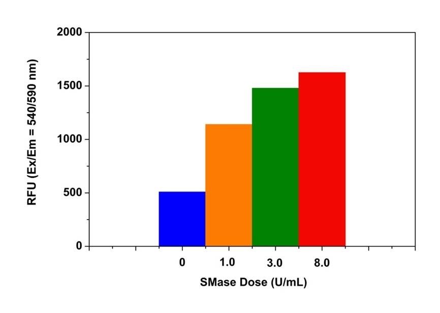
Sphingomyelinase (SMase) is an enzyme that is responsible for cleaving sphingomyelin (SM) to phosphocholine and ceramide. Activation of SMases plays an important role in the cellular response such as regulation of cell growth, cell differentiation, cell cycle arrest and programmed cell death. Five types of sphingomyelinase (SMase) have been identified based on their cation dependence and pH optima of action, including lysosomal acid SMase, secreted zinc-dependent acid SMase, magnesium-dependent neutral SMase, magnesium-independent neutral SMase and alkaline SMase. Among them, the lysosomal acidic SMase and the magnesium-dependent neutral SMase are considered to be the major factors for the production of ceramide in cellular stress responses. Our Amplite® Fluorimetric Acidic Sphingomyelinase Assay Kit provides one of the most sensitive methods for detecting acidic SMase activity or screening its inhibitors. The kit uses Amplite® Red as a fluorogenic probe to indirectly quantify the phosphocholine produced from the hydrolysis of sphingomyelin (SM) by sphingomyelinase (SMase). It can be used for measuring the SMase activity in blood, cell extracts or other solutions. The fluorescence intensity of Amplite® Red is proportional to the formation of phosphocholine, therefore to the SMase activity. The kit is an optimized "mix and read" assay compatible with HTS liquid handling instruments.

Platform


Fluorescence microplate reader

Excitation540 nm
Emission590 nm
Cutoff570 nm
Recommended plateSolid black

Components


Example protocol


AT A GLANCE

Protocol Summary
  1. Prepare acidic SMase standards or SMase test samples (50 µL)
  2. Add sphingomyelin working solution (50 µL)
  3. Incubate at 37 °C for 2 - 3 hours
  4. Add sphingomyelinase working solution (50 µL)
  5. Incubate at RT for 1 - 2 hours
  6. Monitor fluorescence increase at Ex/Em = 540/590 nm (cut off at 570 nm) 
Important      Thaw 1 vial (or bottle) of each kit component to room temperature before starting your experiment.

PREPARATION OF STOCK SOLUTIONS

Unless otherwise noted, all unused stock solutions should be divided into single-use aliquots and stored at -20 °C after preparation. Avoid repeated freeze-thaw cycles.

Amplite™ Red stock solution (200X)
Add 80 µL of DMSO (Component F) into the vial of Amplite™ Red (Component C) to make 200X Amplite™ Red stock solution.
Note     Note: The Amplite™ Red is unstable in the presence of thiols (such as DTT and 2-mercaptoethanol). The final concentration of DTT or 2-mercaptoethanol in the reaction should be lower than 10 µM. Amplite™ Red is also unstable at high pH (>8.5). The reactions should be performed at pH 7 – 8. The assay buffer at pH 7.4 is recommended.

PREPARATION OF STANDARD SOLUTION

For convenience, use the Serial Dilution Planner:
https://www.aatbio.com/tools/serial-dilution/13622


Acidic sphingomyelinase standard
Dilute acidic sphingomyelinase stock solution in 20 mM sodium acetate buffer (pH = 5.0, not provided in the kit). We recommend the concentration range from 10 U/mL to 0.5 U/mL. Note: Acidic sphingomyelinase standard (from human placenta) can be obtained from Sigma-Aldrich (S-5383). Diluted acidic sphingomyelinase standard solution is unstable, and should be used within 4 hours.

PREPARATION OF WORKING SOLUTION

1. Sphingomyelin working solution
Add 50 µL of Sphingomyelin (Component B) to 5 mL of SMase Reaction Buffer (Component D) and mix well.
Note     The sphingomyelin working solution should be used promptly.


2. Sphingomyelinase working solution
Add 5 mL of Assay Buffer (Component E) to the bottle of Enzyme Mix (Component A) and mix well. Add 25 µL of 200X Amplite™ Red stock solution into the bottle of Enzyme Mix solution to make the sphingomyelinase working solution before starting the assay.
Note     The sphingomyelinase working solution should be used promptly and kept from light; longer storage is likely to cause a higher assay background.

SAMPLE EXPERIMENTAL PROTOCOL

Table 1. Layout of acidic sphingomyelinase standards and test samples in a solid black 96-well microplate. SMase = Acidic Sphingomyelinase Standards (SMase1-SMase7, 0.5 to 10 U/mL); BL = Blank Control; TS = Test Samples.
BLBLTSTS
SMase1SMase1......
SMase2SMase2......
SMase3SMase3
SMase4SMase4
SMase5SMase5
SMase6SMase6
SMase7SMase7
Table 2. Reagent composition for each well
WellVolumeReagent
SMase1 - SMase7 50 µL Serial Dilution (0.5 to 10 U/mL)
BL 50 µL 20 mM Sodium Acetate Buffer (pH = 5)
TS 50 µL Test Sample
  1. Add the acidic sphingomyelinase standards and sphingomyelinase-containing test samples into a solid black 96-well microplate as shown in Tables 1 and 2. For a 384-well plate, use 25 µL of reagent per well instead of 50 µL.
    Note     Treat your cells or tissue samples as desired.
  2. Add 50 µL of sphingomyelin working solution into each well of the sphingomyelinase standards, blank control and test samples. Add the diluted acidic sphingomyelinase standards in duplicate. For a 384-well plate, add 25 µL of sphingomyelin working solution into each well instead, for a total volume of 50 µL/well.
  3. Incubate the reaction mixture at 37 °C for 2 - 3 hours.
  4. Add 50 µL of sphingomyelinase working solution into each well of the acidic sphingomyelinase standards, blank control, and test samples to make the total sphingomyelinase assay volume of 150 µL/well. For a 384-well plate, add 25 µL sphingomyelinase assay working solution into each well instead, for the total sphingomyelin assay volume of 75 µL/well.
  5. Incubate the enzyme reaction mixture for 1 - 2 hours at room temperature (protected from light).
  6. Monitor the fluorescence increase with a fluorescence microplate reader at Ex/Em = 540/590 nm (cut off at 570 nm). 

Spectrum


Open in Advanced Spectrum Viewer
spectrum

Spectral properties

Excitation (nm)571
Emission (nm)584

Images


Citations


View all 9 citations: Citation Explorer
Extracellular high molecular weight $\alpha$-synuclein oligomers induce cell death by disrupting the plasma membrane
Authors: Ito, Naohito and Tsuji, Mayumi and Adachi, Naoki and Nakamura, Shiro and Sarkar, Avijite Kumer and Ikenaka, Kensuke and Aguirre, C{\'e}sar and Kimura, Atsushi Michael and Kiuchi, Yuji and Mochizuki, Hideki and others,
Journal: npj Parkinson's Disease (2023): 139
Sortilin deletion in the prefrontal cortex and hippocampus ameliorates depressive-like behaviors in mice via regulating ASM/ceramide signaling
Authors: Chen, Shu-jian and Gao, Cong-cong and Lv, Qun-yu and Zhao, Meng-qi and Qin, Xiao-ying and Liao, Hong
Journal: Acta Pharmacologica Sinica (2021): 1--15
Doxepin Mitigates Noise-induced Neuronal Damage in Primary Auditory Cortex of Mice via Suppression of Acid Sphingomyelinase/Ceramide Pathway
Authors: Su, Yu-Ting and Meng, Xing-Xing and Zhang, Xi and Guo, Yi-Bin and Zhang, Hai-Jun and Cheng, Yao-Ping and Xie, Xiao-Ping and Chang, Yao-Ming and Bao, Jun-Xiang
Journal: The Anatomical Record (2017): 2220--2232
Doxepin mitigates noise induced neuronal damage in primary auditory cortex of mice via suppression of acid sphingomyelinase/ceramide pathway
Authors: Su, Yu-Ting and Meng, Xing-Xing and Zhang, Xi and Guo, Yi-Bin and Zhang, Hai-Jun and Cheng, Yao-Ping and Xie, Xiao-Ping and Chang, Yao-Ming and Bao, Jun-Xiang
Journal: The Anatomical Record (2017)
Riccardin DN induces lysosomal membrane permeabilization by inhibiting acid sphingomyelinase and interfering with sphingomyelin metabolism in vivo
Authors: Li, Lin and Niu, Huanmin and Sun, Bin and Xiao, Yanan and Li, Wei and Yuan, Huiqing and Lou, Hongxiang
Journal: Toxicology and Applied Pharmacology (2016): 175--184
New Aspects of Silibinin Stereoisomers and their 3-O-galloyl Derivatives on Cytotoxicity and Ceramide Metabolism in Hep G2 hepatocarcinoma Cell Line
Authors: Mashhadi Akbar Boojar, Mahdi and Ejtemaei Mehr, Shahram and Hassanipour, Mahsa and Mashhadi Akbar Boojar, Masoud and Dehpour, Ahmad Reza
Journal: Iranian Journal of Pharmaceutical Research (2016): 421--433
Mitochondrial respiration controls lysosomal function during inflammatory T cell responses
Authors: Baixauli, Francesc and Acín-Pérez, Rebeca and Villarroya-Beltrí, Carolina and Mazzeo, Carla and Nunez-Andrade, Norman and Gab, undefined and é-Rodriguez, Enrique and Ledesma, Maria Dolores and Blázquez, Alberto and Martin, Miguel Angel and Falcón-Pérez, Juan Manuel and others, undefined
Journal: Cell metabolism (2015): 485--498
The ATP-binding cassette transporter-2 (ABCA2) regulates esterification of plasma membrane cholesterol by modulation of sphingolipid metabolism
Authors: Davis, Warren
Journal: Biochimica et Biophysica Acta (BBA)-Molecular and Cell Biology of Lipids (2014): 168--179
A high-throughput sphingomyelinase assay using natural substrate
Authors: Xu, Miao and Liu, Ke and Southall, Noel and Marugan, Juan J and Remaley, Alan T and Zheng, Wei
Journal: Analytical and bioanalytical chemistry (2012): 407--414

References


View all 65 references: Citation Explorer
Phospholipase C and sphingomyelinase activities of the Clostridium perfringens alpha-toxin
Authors: Urbina P, Flores-Diaz M, Alape-Giron A, Alonso A, Goni FM.
Journal: Chem Phys Lipids (2009): 51
Sphingomyelinase-induced ceramide production stimulate calcium-independent JNK and PP2A activation following cerebral ischemia
Authors: Tian HP, Qiu TZ, Zhao J, Li LX, Guo J.
Journal: Brain Inj (2009): 1073
SMase II, a new sphingomyelinase D from Loxosceles laeta venom gland: molecular cloning, expression, function and structural analysis
Authors: de Santi Ferrara GI, Fern and es-Pedrosa Mde F, Junqueira-de-Azevedo Ide L, Goncalves-de-Andrade RM, Portaro FC, Manzoni-de-Almeida D, Murakami MT, Arni RK, van den Berg CW, Ho PL, Tambourgi DV.
Journal: Toxicon (2009): 743
Neutral sphingomyelinase-induced ceramide accumulation by oxidative stress during carbon tetrachloride intoxication
Authors: Ichi I, Kamikawa C, Nakagawa T, Kobayashi K, Kataoka R, Nagata E, Kitamura Y, Nakazaki C, Matsura T, Kojo S.
Journal: Toxicology (2009): 33
Purification, characterization, and gene cloning of sphingomyelinase C from Streptomyces griseocarneus NBRC13471
Authors: Sugimori D., undefined
Journal: J Biosci Bioeng (2009): 293
Transcriptional regulation of neutral sphingomyelinase 2 gene expression of a human breast cancer cell line, MCF-7, induced by the anti-cancer drug, daunorubicin
Authors: Ito H, Murakami M, Furuhata A, Gao S, Yoshida K, Sobue S, Hagiwara K, Takagi A, Kojima T, Suzuki M, Banno Y, Tanaka K, Tamiya-Koizumi K, Kyogashima M, Nozawa Y, Murate T.
Journal: Biochim Biophys Acta (2009): 681
ApoCIII-enriched LDL in type 2 diabetes displays altered lipid composition, increased susceptibility for sphingomyelinase, and increased binding to biglycan
Authors: Hiukka A, Stahlman M, Pettersson C, Levin M, Adiels M, Teneberg S, Leinonen ES, Hulten LM, Wiklund O, Oresic M, Olofsson SO, Taskinen MR, Ekroos K, Boren J.
Journal: Diabetes (2009): 2018
Structure and function of sphingomyelinase
Authors: Oda M., undefined
Journal: Yakugaku Zasshi (2009): 1233
Expression of alkaline sphingomyelinase in yeast cells and anti-inflammatory effects of the expressed enzyme in a rat colitis model
Authors: Andersson D, Kotarsky K, Wu J, Agace W, Duan RD.
Journal: Dig Dis Sci (2009): 1440
A complex extracellular sphingomyelinase of Pseudomonas aeruginosa inhibits angiogenesis by selective cytotoxicity to endothelial cells
Authors: Vasil ML, Stonehouse MJ, Vasil AI, Wadsworth SJ, Goldfine H, Bolcome RE, 3rd, Chan J.
Journal: PLoS Pathog (2009): e1000420