Products
Services
Resources
Selection Guides
About
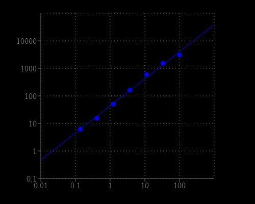
Amplite® Fluorimetric α-Ketoglutarate Quantitation Kit
Alpha-ketoglutarate (α-ketoglutarate) is a key molecule in the Krebs cycle determining the overall rate of the citric acid cycle of the organism. As a precursor of glutamate and glutamine, α-ketoglutarate is a central metabolic fuel for cells of the gastrointestinal tract as well. It can decrease protein catabolism and increase protein synthesis to enhance bone tissue formation in the skeletal muscles and can be used in clinical applications. Alpha-ketoglutarate is used for kidney disease; intestinal and stomach disorders, including bacterial infections; liver problems; cataracts; and recurring yeast infections. It is also used for improving the way kidney patients receiving hemodialysis treatments process protein. Amplite®™ Fluorimetric α-Ketoglutarate Quantitation Kit offers a sensitive fluorimetric assay for quantifying α-ketoglutarate in biological samples. It utilizes an enzyme coupled reaction that releases hydrogen peroxide, which can be detected with Amplite®™ Red at Ex/Em ~540/590 nm (red fluorescence).
| Catalog | Size | Price | Quantity |
|---|---|---|---|
| 10087 | 200 Tests | Price |
Storage, safety and handling
| H-phrase | H303, H313, H333 |
| Hazard symbol | XN |
| Intended use | Research Use Only (RUO) |
| R-phrase | R20, R21, R22 |
| UNSPSC | 12171501 |
Instrument settings
| Fluorescence microplate reader | |
| Excitation | 540 nm |
| Emission | 590 nm |
| Cutoff | 570 nm |
| Recommended plate | Solid black |
Contact us
| Telephone | |
| Fax | |
| sales@aatbio.com | |
| International | See distributors |
| Bulk request | Inquire |
| Custom size | Inquire |
| Technical Support | Contact us |
| Request quotation | Request |
| Purchase order | Send to sales@aatbio.com |
| Shipping | Standard overnight for United States, inquire for international |
Page updated on March 25, 2026