logo
AAT Bioquest

Amplite® Fluorimetric Beta-Galactosidase Assay Kit *Green Fluorescence*

β-galactosidase dose response was measured with Amplite™ Fluorimetric beta-Galactosidase Assay Kit in a Costar solid black 96-well plate using Gemini fluorescence microplate reader (Molecular Devices). 
Ordering information
Price
Catalog Number
Unit Size
Quantity
Add to cart
Additional ordering information
Telephone1-800-990-8053
Fax1-800-609-2943
Emailsales@aatbio.com
InternationalSee distributors
Bulk requestInquire
Custom sizeInquire
ShippingStandard overnight for United States, inquire for international
Request quotation
Spectral properties
Absorbance (nm)487
Correction Factor (260 nm)0.32
Correction Factor (280 nm)0.35
Extinction coefficient (cm -1 M -1)800001
Excitation (nm)498
Emission (nm)517
Quantum yield0.79001, 0.952
Storage, safety and handling
H-phraseH303, H313, H333
Hazard symbolXN
Intended useResearch Use Only (RUO)
R-phraseR20, R21, R22
UNSPSC12352200

OverviewpdfSDSpdfProtocol


Absorbance (nm)
487
Correction Factor (260 nm)
0.32
Correction Factor (280 nm)
0.35
Extinction coefficient (cm -1 M -1)
800001
Excitation (nm)
498
Emission (nm)
517
Quantum yield
0.79001, 0.952
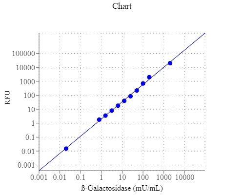
E. coli beta-galactosidase is a 464 kD tetramer. Each unit of beta-galactosidase consists of five domains, the third of which is the active site. It is an essential enzyme in cells. Deficiencies in this enzyme can result in galactosialidosis or Morquio B syndrome. In E. coli, beta-galactosidase is produced by the activation of LacZ operon. Detection of LacZ expression has become routine to the point of detection of as few as 5 copies of ?-galactosidase per cell. This kit uses a fluorogenic galactosidase substrate that can sensitively distinguish LacZ+ vs. LacZ-cells. It can be used either for detecting galactosidase conjugates in ELISA type assay systems or for monitoring LacZ gene expression in cells. The galactosidase-cleaved product has an emission spectra that can be detected with most of fluorescence instruments equipped with a FITC filter set.

Platform


Fluorescence microplate reader

Excitation490 nm
Emission525 nm
Cutoff515 nm
Recommended plateSolid black

Components


Example protocol


AT A GLANCE

Protocol Summary
  1. Prepare stable or transient transfected cells with LacZ gene
  2. Incubate cells (samples) with test compounds
  3. Lyse the cells
  4. Transfer the lysate to a microtiter plate
  5. Add FDG working solution
  6. Incubate at room temperature or 37°C for at least 5 minutes depending on cell type
  7. Add stopping solution
  8. Monitor fluorescence intensity at Ex/Em = 490/525 nm 
Important      Thaw all the kit components to room temperature before use.

CELL PREPARATION

For guidelines on cell sample preparation, please visit https://www.aatbio.com/resources/guides/cell-sample-preparation.html

PREPARATION OF STOCK SOLUTIONS

Unless otherwise noted, all unused stock solutions should be divided into single-use aliquots and stored at -20 °C after preparation. Avoid repeated freeze-thaw cycles.

FDG stock solution (1X)
Add 125 µL of DMSO (Component E) into the vial of FDG (Component A) to make 1X FDG stock solution. Note: 25 µL of FDG is enough for 1 plate. Keep from light.

PREPARATION OF STANDARD SOLUTION

For convenience, use the Serial Dilution Planner:
https://www.aatbio.com/tools/serial-dilution/12601


β-Galactosidase standard
Optional (if a standard curve is desired): Prepare a serial dilution of β-galactosidase (E. Coli) standards with 0.3% β- mercaptoethanol assay buffer. Transfer 50 µL aliquot of each point on the standard curve to the control wells of the plate. The highest recommended amount of β-galactosidase is 200 mU/mL (200 - 400 ng). 2X serial dilution of standard curve consisting of 8 points is recommended. Note: Adjust the standard curve to suit the specific experimental conditions, such as cell type, number, transfection effeciency, and size of the culture plates. The dilutions for the standard curve must be prepared freshly each time the assay is performed.

PREPARATION OF WORKING SOLUTION

1. 0.3 % β-mercaptoethanol assay buffer
Add 30 µL of β-mercaptoethanol (Component F) to 10 mL of Reaction Buffer (Component B), and mix well. Note: Additional buffer is needed for preparing enzyme dilution buffer, which is used to generate a standard curve.

2. FDG working solution
Add 25 µL of 1X FDG stock solution into 5 mL of 0.3 % β-mercaptoethanol assay buffer. Note: DO NOT keep FDG solutions at room temperature for an extended period of time as spontaneous hydrolysis will occur.

3. Lysis buffer working solution
Add 5 µL of β-mercaptoethanol (Component F) to 5 mL of Lysis Buffer (Component D) before use. Note: Always add 0.1% β-mercaptoethanol into lysis buffer before lysing the cells

SAMPLE EXPERIMENTAL PROTOCOL

Table 1. Recommended Lysis Buffer working solution volumes for cell culture plates.
Type of culture platesLysis Buffer working solutions (µL/well)
96-well plate50
24-well plate250
12-well plate500
6-well plate1000
60 mm plate2000
100 mm plate4000

Prepare cell extracts from mammalian cells
  1. Treat cells containing LacZ gene with test compounds for a desired period of time.
  2. Wash the cells twice with 1X PBS. Do not dislodge the cells.
  3. Lyse cells accordingly with Lysis Buffer working solution.For adherent cells: Add Lysis Buffer working solution to the culture plates. See table 1 for recommended volumes.For non-adherent cells: Pellet the cells into centrifuge tube, and add 50 - 2000 µL (depending on the size of the cell pellet) of Lysis Buffer working solution to the tube.
  4. Incubate cells from previous step at room temperature for 10 - 15 minutes, and gently swirl the plates or tubes several times to ensure complete lysis.
  5. Proceed to the FDG assay or freeze the sample at -80 °C until use. Note: A good lysis can also be obtained by a quick freeze-and-thaw cycle (freeze 1 - 2 hours at -20°C to -80°C and thaw at room temperature). Alternatively, centrifuge the cell lysis for 2 - 3 minutes to pellet the insoluble material, and then assay the supernatant. 

Run ß-galactosidase assay
  1. Thaw the tube or plate of lysed cells at room temperature if needed. Perform the assay directly on the 96-well plate if the cells were seeded in a 96-well plate.
  2. Add 50 µL of cell extracts into each well of the 96-well plate. Save some control wells for the standard curve (50 µL/well) if a standard curve is desired. Note: If necessary, dilute the lysate in Lysis Buffer working solution when transfection efficiency is very high or reduce the volume of lysis buffer when transfection efficiency is low. If the transfection is performed in a 96-well plate, or a stable cell line was seeded into a 96-well plate, perform the assay directly on the plate. For endogenous β-galactosidase activity control, add 50 µL of cell lysate from non-transfected cells. For blank control, add 50 µL of 1X lysis buffer.
  3. Add 50 µL of FDG working solution to each well. Incubate the plate at room temperature or 37°C for approximately 5 min to 4 hr depending on the cell type.
  4. Add 50 µL of Stop Buffer (Component C) to each well. The stop buffer causes an increase in the fluorescence intensity of the product, in addition to terminate the reaction.
  5. Measure the fluorescence intensity of the solution in each well with a fluorescence microplate reader at Ex/Em = 490/525 nm.
  6. Quantify ß-galactosidase expression based on a linear standard curve. 

Spectrum


Open in Advanced Spectrum Viewer
spectrum

Spectral properties

Absorbance (nm)487
Correction Factor (260 nm)0.32
Correction Factor (280 nm)0.35
Extinction coefficient (cm -1 M -1)800001
Excitation (nm)498
Emission (nm)517
Quantum yield0.79001, 0.952

Product Family


NameExcitation (nm)Emission (nm)Extinction coefficient (cm -1 M -1)Quantum yield
Amplite® Fluorimetric Beta-Galactosidase Assay Kit *Red Fluorescence*5715846500010.751

Images


Citations


View all 1 citations: Citation Explorer
BZLF1 Attenuates Transmission of Inflammatory Paracrine Senescence in Epstein-Barr Virus-Infected Cells by Downregulating Tumor Necrosis Factor Alpha
Authors: Long, Xubing and Li, Yuqing and Yang, Mengtian and Huang, Lu and Gong, Weijie and Kuang, Ersheng
Journal: Journal of Virology (2016): 7880--7893

References


View all 104 references: Citation Explorer
One-step high-throughput assay for quantitative detection of beta-galactosidase activity in intact gram-negative bacteria, yeast, and mammalian cells
Authors: Vidal-Aroca F, Giannattasio M, Brunelli E, Vezzoli A, Plevani P, Muzi-Falconi M, Bertoni G.
Journal: Biotechniques (2006): 433
A homogeneous cell-based assay to measure nuclear translocation using beta-galactosidase enzyme fragment complementation
Authors: Fung P, Peng K, Kobel P, Dotimas H, Kauffman L, Olson K, Eglen RM.
Journal: Assay Drug Dev Technol (2006): 263
Microfluidic sequential injection analysis in a short capillary
Authors: Du WB, Fang Q, Fang ZL.
Journal: Anal Chem (2006): 6404
Use of monoclonal antibodies to quantify the dynamics of alpha-galactosidase and endo-1,4-beta-glucanase production by Trichoderma hamatum during saprotrophic growth and sporulation in peat
Authors: Thornton CR., undefined
Journal: Environ Microbiol (2005): 737
Identification of bacteria with beta-galactosidase activity in faeces from lactase non-persistent subjects
Authors: He T, Priebe MG, Vonk RJ, Welling GW.
Journal: FEMS Microbiol Ecol (2005): 463
Translocation of beta-galactosidase mediated by the cell-penetrating peptide pep-1 into lipid vesicles and human HeLa cells is driven by membrane electrostatic potential
Authors: Henriques ST, Costa J, Castanho MA.
Journal: Biochemistry (2005): 10189
High-throughput screening of enzyme libraries: in vitro evolution of a beta-galactosidase by fluorescence-activated sorting of double emulsions
Authors: Mastrobattista E, Taly V, Chanudet E, Treacy P, Kelly BT, Griffiths AD.
Journal: Chem Biol (2005): 1291
Cellular logic with orthogonal ribosomes
Authors: Rackham O, Chin JW.
Journal: J Am Chem Soc (2005): 17584
New transport peptides broaden the horizon of applications for peptidic pharmaceuticals
Authors: Langedijk JP, Olijhoek T, Schut D, Autar R, Meloen RH.
Journal: Mol Divers (2004): 101
In vivo imaging of beta-galactosidase activity using far red fluorescent switch
Authors: Tung CH, Zeng Q, Shah K, Kim DE, Schellingerhout D, Weissleder R.
Journal: Cancer Res (2004): 1579