logo
AAT Bioquest

Amplite® Fluorimetric DPP4 Activity Assay Kit

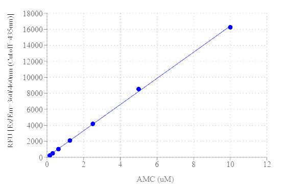
The Amplite® Fluorimetric DPP4 Activity Assay Kit provides a simple, quick, and direct protocol for measuring DPP4 Activity. This assay leverages the DPP4-mediated cleavage of a non-fluorescent substrate, resulting in a fluorescent product with Ex/Em = 360/460 nm, respectively. The fluorescence intensity generated is directly proportional to the DPP4 activity in the sample, making it suitable for detecting DPP4 in various biological matrices such as cellular lysates, tissue extracts, and serum. Additionally, the assay is compatible with high-throughput screening systems. One unit (U) is the amount of enzyme that catalyzes the reaction of 1 µmol of substrate per minute. Dipeptidyl peptidase-4, also known as DPP4, CD26, ADCP2, DPP, is a transmembrane glycoprotein belonging to the prolyl oligopeptidase family. It is a serine exopeptidase that cleaves X-proline and X-Alanine residues from the N-terminal ends of polypeptides. DPP4 is involved in several physiological processes, including glucose metabolism through the regulation of glucagon-like-peptide (GLP-1), immune regulation by acting as a receptor on many immune cells, signal transduction as a transmembrane protein responsive to growth factors and chemokines, and tumor suppression via immune modulation. DPP4 inhibitors are being used as a treatment for type-2 diabetes.

Example protocol

AT A GLANCE

Important Note

Thaw all the kit components at room temperature before starting the experiment.

Protocol Summary
  1. Prepare the test samples, DPP4 Positive Control, and the serially diluted AMC standards (50 μL).

  2. Add the DPP4 working solution (50 μL).

  3. Incubate at room temperature for 10-30 minutes.

  4. Monitor the fluorescence intensity at Ex/Em=360/460 nm, cutoff=435 nm.

PREPARATION OF STANDARD SOLUTIONS

For convenience, use the Serial Dilution Planner:
https://www.aatbio.com/tools/serial-dilution/11323

AMC Standard
Add 5 μL of 1 mM AMC standard solution to 495 μL of DPP4 Assay Buffer (Component A) to prepare a 10 μM AMC standard solution (STD7). Then, take 300 μL of STD7 and perform 1:2 serial dilutions in of DPP4 Assay Buffer (Component A) to create a series of AMC standards from STD7 to STD1.

PREPARATION OF WORKING SOLUTION

DPP4 Working Solution
  1. Add 0.25 mL of the DPP4 Substrate (Component B) to 5 mL of the DPP4 Assay Buffer (Component A), and mix well.

    Note: This DPP4 working solution should be freshly prepared before each experiment and protected from light. A 5 mL solution is enough for 100 tests. Please prepare the necessary amount of DPP4 working solution based on this proportion.

5X DPP4 Inhibitor Working Solution
  1. Add 2.5 µL of DPP4 Inhibitor (Sitagliptin) (Component E) to 500 µL of DPP4 Assay Buffer (Component A) to create a 5X DPP4 Inhibitor Working Solution.

DPP4 Positive Control
  1. Prepare one or more DPP4 positive controls along with your test sample. The suggested concentration for these controls is between 0.5 µg/mL and 0.05 µg/mL in the DPP4 Assay Buffer (Component A). For example, to make a 0.5 µg/mL DPP4 positive control, add 2.5 µL of the DPP4 Positive Control Stock Solution to 250 µL of the DPP4 Assay Buffer (Component A).

Test Samples
  1. Tissue and cells can be homogenized in the DPP4 Assay Buffer (Component A). After homogenization, centrifuge the sample at 13,000xg for 10 minutes to remove insoluble material. Serum samples can be directly added to the wells. Adjust the final volume of all samples to 50 µL using DPP4 Assay Buffer.

SAMPLE EXPERIMENTAL PROTOCOL

Table 1. Layout of AMC standards and test samples in a 96-well clear bottom microplate. (STD = AMC Standards (STD7-STD1, 10 to 0.0781 µM), BL= Blank Control, TS = Test Samples.)

BL
BL
Positive Control
Inhibitor Control
TS
STD 1
STD 1
...
...
...
STD 2
STD 2
...
...
...
STD 3
STD 3
STD 4
STD 4
STD 5
STD 5
STD 6
STD 6
STD 7
STD 7

Table 2. Reagent composition for each well.

Well
Volume
Reagent
STD 1 -STD 7
50 µL
AMC Serial Dilutions (0.0781-10 µM)
BL
50 µL
DPP4 Assay buffer
DPP4 Positive Control
50 µL
40 µL DPP4 Positive Control + 10 µL DPP4 Assay buffer
DPP4 Inhibitor Control
50 µL
40 µL DPP4 Positive Control + 10 µL 5X DPP4 Inhibitor Working Solution
TS
50 µL
Cell content
  1. Prepare the AMC standards (STD1-7), blank controls (BL), DPP4 Positive Control, DPP4 Inhibitor Control, and test samples (TS) according to the layout provided in Tables 1 and 2. When using a 384-well plate, use 25 µL of reagent per well instead of 50 µL.

  2. Add 50 µL of DPP4 Working Solution to each well containing the blank control, DPP4 Positive Control, DPP4 Inhibitor Control, and test samples . For a 384-well plate, add 25 µL of DPP4 Working Solution to each well instead.

  3. Incubate at room temperature for 10–30 minutes, protected from light.

  4. Monitor the fluorescence intensity with a fluorescence microplate reader at Ex/Em = 360/460 nm, Cutoff = 435 nm.

References

View all 50 references: Citation Explorer
Dipeptidyl-peptidase 4 (DPP4) mediates fatty acid uptake inhibition by glucose via TAS1R3 and GLUT-2 in Caco-2 enterocytes.
Authors: Preinfalk, Verena and Kimmeswenger, Isabella and Somoza, Veronika and Lieder, Barbara
Journal: Heliyon (2024): e30329
Diabetic individuals with COVID-19 exhibit reduced efficacy of gliptins in inhibiting dipeptidyl peptidase 4 (DPP4). A suggested explanation for increased COVID-19 susceptibility in patients with type 2 diabetes mellitus (T2DM).
Authors: Mora-Rodríguez, José María and Sánchez, Belén G and Bort, Alicia and Díaz-Yuste, Alba and Ballester-González, Rubén and Arrieta, Francisco and Sebastián-Martín, Alba and Díaz-Laviada, Inés
Journal: Life sciences (2024): 122292
Increased Thyroid DPP4 Expression Is Associated With Inflammatory Process in Patients With Hashimoto Thyroiditis.
Authors: Wen, Xiaohui and Chang, Xiaona and He, Xueqing and Cai, Qingyun and Wang, Guang and Liu, Jia
Journal: The Journal of clinical endocrinology and metabolism (2024): 1517-1525
Bidirectional relation between dipeptidyl peptidase 4 and angiotensin II type I receptor signaling.
Authors: Martins, Flavia L and Ribeiro-Silva, Joao Carlos and Nistala, Ravi and Girardi, Adriana C C
Journal: American journal of physiology. Cell physiology (2024): C1203-C1211
Pyrano[2,3-c]pyrazole fused spirooxindole-linked 1,2,3-triazoles as antioxidant agents: Exploring their utility in the development of antidiabetic drugs via inhibition of α-amylase and DPP4 activity.
Authors: Chahal, Sandhya and Rani, Payal and Shweta, and Goel, Kapil Kumar and Joshi, Gaurav and Singh, Rajvir and Kumar, Parvin and Singh, Devender and Sindhu, Jayant
Journal: Bioorganic chemistry (2024): 107363
Page updated on April 25, 2025

Ordering information

Price
Unit size
Catalog Number11323
Quantity
Add to cart

Additional ordering information

Telephone1-800-990-8053
Fax1-800-609-2943
Emailsales@aatbio.com
InternationalSee distributors
Bulk requestInquire
Custom sizeInquire
Technical SupportContact us
Purchase orderSend to sales@aatbio.com
ShippingStandard overnight for United States, inquire for international
Request quotation

Storage, safety and handling

H-phraseH303, H313, H333
Hazard symbolXN
Intended useResearch Use Only (RUO)
R-phraseR20, R21, R22
UNSPSC12352200

Platform

Fluorescence microplate reader

Excitation360 nm
Emission460 nm
Cutoff435 nm
Recommended plateSolid black or black plate with clear bottom

Components

AMC dose response was measured with the Amplite® Fluorimetric DPP4 Activity Assay Kit on a 96-well black microplate after incubation for 30 min at room temperature. The fluorescence intensity was monitored using a Gemini fluorescence microplate reader (Molecular Devices) Ex/Em = 360/460 nm, Cutoff = 435 nm.