Products
Services
Resources
Selection Guides
About
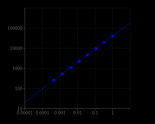
Amplite® Fluorimetric Endotoxin Detection Kit
Lipopolysaccharide (LPS), also known as endotoxin, is the major component of the outer membrane of Gram-negative bacteria. LPS is a potent stimulator of the vertebrate innate immune system and can cause fever, septic shock and eventually death. LPS is also recognized as a biomarker for the detection of bacterial pathogen invasion, and is responsible for the development of inflammatory response and endotoxic shock in extreme cases. Detection of LPS in biological materials, such as protein, peptide or antibody sample, is a critical task in biological manufacturing and processing. Amplite® Fluorimetric Endotoxin Detection Kit uses Endotoxin Green™, a sensitive fluorogenic substrate. Endotoxin Green™ is hydrolyzed in the presence of endotoxins and the Limulus Amebocyte Lysate (LAL), an extract of blood cells from a horseshoe crab. The hydrolyzed product of Endotoxin Green™ generates strong green fluorescence. The endotoxin activity is proportional to the fluorescence intensity resulted from the hydrolysis of Endotoxin Green™. Amplite® Fluorimetric Endotoxin Detection Kit can detect a broad range of endotoxin (from 1 EU/ml to 0.001 EU/ml). It is very sensitive and can detect as low as 0.001 EU/mL of endotoxin within 30 min.
| Catalog | Size | Price | Quantity |
|---|---|---|---|
| 60006 | 100 Tests | Price |
Storage, safety and handling
| H-phrase | H303, H313, H333 |
| Hazard symbol | XN |
| Intended use | Research Use Only (RUO) |
| R-phrase | R20, R21, R22 |
| UNSPSC | 12171501 |
Instrument settings
| Fluorescence microplate reader | |
| Excitation | 490 nm |
| Emission | 525 nm |
| Cutoff | 515 nm |
| Recommended plate | Solid black |
| Instrument specification(s) | Top read mode |
Contact us
| Telephone | |
| Fax | |
| sales@aatbio.com | |
| International | See distributors |
| Bulk request | Inquire |
| Custom size | Inquire |
| Technical Support | Contact us |
| Request quotation | Request |
| Purchase order | Send to sales@aatbio.com |
| Shipping | Standard overnight for United States, inquire for international |
Page updated on May 16, 2026