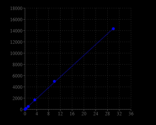
Amplite® Fluorimetric Glucose Quantitation Kit
Price | |
Catalog Number | |
Unit Size | |
Quantity |
Telephone | 1-800-990-8053 |
Fax | 1-800-609-2943 |
sales@aatbio.com | |
Quotation | Request |
International | See distributors |
Shipping | Standard overnight for United States, inquire for international |
Excitation (nm) | 571 |
Emission (nm) | 584 |
H-phrase | H303, H313, H333 |
Hazard symbol | XN |
Intended use | Research Use Only (RUO) |
R-phrase | R20, R21, R22 |
UNSPSC | 12352200 |
Overview | ![]() ![]() |
Excitation (nm) 571 | Emission (nm) 584 |
Platform
Fluorescence microplate reader
Excitation | 540 nm |
Emission | 590 nm |
Cutoff | 570 nm |
Recommended plate | Solid black |
Components
Example protocol
AT A GLANCE
Protocol summary
- Prepare and add Glucose standards and/or test samples (50 µL)
- Prepare and add Glucose Assay working solution (50 µL)
- Incubate at 37°C for 10 - 30 minutes
- Monitor fluroscence intensity at Ex/Em = 540/590 nm
Important notes
Thaw all the kit components at room temperature before starting the experiment.
PREPARATION OF STOCK SOLUTION
1. Amplite™ Red stock solution (250X):
Add 100 µL of DMSO (Component E) into the vial of Amplite™ Red substrate (Component A). The stock solution should be used promptly. Any remaining solution should be aliquoted and refrozen at -20 oC. Note: Avoid repeated freeze-thaw cycles. Note: The Amplite™ Red substrate is unstable in the presence of thiols such as dithiothreitol (DTT) and 2-mercaptoethanol. The final concentration of DTT or 2-mercaptoethanol in the reaction should be no higher than 10 µM. The Amplite™ Red substrate is also unstable at high pH (> 8.5). Therefore, the reaction should be performed at pH 7–8. The provided assay buffer (pH 7.4) is recommended.
2. Horseradish Peroxidase (HRP) stock solution (10 U/mL):
Add 1 mL of Assay Buffer (Component B) into the vial of Horseradish Peroxidase (Component C). Note: The unused HRP solution should be divided into single use aliquotes and stored at -20 oC.
3. Glucose Oxidase solution (100 U/mL):
Add 1 mL of Assay Buffer (Component B) into the vial of Glucose Oxidase (Component D). Note: The unused Glucose Oxidase solution should be divided into single use aliquotes and stored at -20 oC.
4. Glucose stock solution (800mM):
Add 1 mL of Assay Buffer (Component B) into the vial of Glucose (Component F). Note: The unused Glucose solution should be divided into single use aliquotes and stored at -20 oC.
PREPARATION OF STANDARD SOLUTION
For convenience, use the Serial Dilution Planner: https://www.aatbio.com/tools/serial-dilution/40005
Prepare a glucose standard by diluting the appropriate amount of the 800 mM glucose stock solution into Assay Buffer (Component B) to produce glucose concentrations of 30 µM. Then perform 1:3 serial dilutions in Assay Buffer (Component B) to get approximately 10, 3, 1, 0.3, 0.1 and 0.03 µM serially diluted glucose standards. A non-glucose buffer control is included as blank control.
PREPARATION OF WORKING SOLUTION
Table 1. Assay working solution for one clear bottom 96-well microplate (2X)
Components |
Volume |
Amplite™ Red Stock Solution (250x) |
20 μL |
HRP Stock Solution (10 U/mL) |
100 μL |
Glucose Oxidase Solution (100 U/mL) |
100 μL |
Assay Buffer |
4.78 mL |
Total volume |
5 mL |
SAMPLE EXPERIMENTAL PROTOCOL
Table 2. Layout of Glucose standards and test samples in a solid black 96-well microplate. GS = Glucose standard (GS1-GS7); BL = blank control; TS = test sample.
BL | BL | TS | TS |
GS1 | GS1 | ... | ... |
GS2 | GS2 | ... | ... |
GS3 | GS3 | ||
GS4 | GS4 | ||
GS5 | GS5 | ||
GS6 | GS6 | ||
GS7 | GS7 |
Table 3. Reagent composition for each well
Glucose Standard | Blank Control | Test Sample |
Serial Dilutions: 50 µL | Assay Buffer (Compound B): 50 µL | 50 µL |
Note: High Concentration of glucose (e.g. 100 µM in test sample or standard) may cause reduced fluoroscence signal due to the overoxidation of Amplite™ red substrate (to a non-fluorescent product).
Glucose assay
- Add glucose standards and glucose containing test samples into a 96-well solid black microplate as described in Tables 2 and 3.
- Add 50 µL of Glucose Assay working solution into each well of glucose standard, blank control, and test samples (Table 2) to make the total glucose assay volume of 100 µL/well. Note: For a 384-well plate, add 25 µL of sample and 25 µL of assay reaction mixture into each well.
- Incubate the reaction for 10 to 30 minutes at 37 oC, protected from light.
- Monitor the fluorescence intensity with a fluorescence plate reader at Ex/Em= 530-570 nm/590-600 nm (optimal Ex/Em = 540/590 nm).
Images
Citations
Authors: Callet, Th{\'e}r{\`e}se and Li, Hongyan and Heraud, C{\'e}cile and Larroquet, Laurence and Lanuque, Anthony and Sandres, Franck and Terrier, Fr{\'e}d{\'e}ric and Surget, Anne and Corraze, Genevi{\`e}ve and Panserat, St{\'e}phane and others,
Journal: animal (2022): 100670
Authors: Jacquier, Valentin and Gitenay, Delphine and Fritsch, Samuel and Bonnet, Sandrine and Gy{\H{o}}rffy, Bal{\'a}zs and Jalaguier, St{\'e}phan and Linares, Laetitia K and Cavaill{\`e}s, Vincent and Teyssier, Catherine
Journal: Cellular and Molecular Life Sciences (2022): 1--17
Authors: Callet, Therese and Li, Hongyan and Surget, Anne and Terrier, Frederic and Sandres, Franck and Lanuque, Anthony and Panserat, Stephane and Marandel, Lucie
Journal: PeerJ (2021): e12102
Authors: Deng, Li-li
Journal: Archives of Medical Research (2020)
Authors: Jacquier, Valentin and Gitenay, Delphine and Fritsch, Samuel and Linares, Laetitia K and Bonnet, Sandrine and Jalaguier, St{\'e}phan and Cavaill{\`e}s, Vincent and Teyssier, Catherine
Journal: bioRxiv (2020)
Authors: Callet, Th{\'e}r{\`e}se and Hu, Huihua and Larroquet, Laurence and Surget, Anne and Liu, Jingwei and Plagnes-Juan, Elisabeth and Maunas, Patrick and Turonnet, Nicolas and Mennigen, Jan Alexander and Bobe, Julien and others,
Journal: Frontiers in physiology (2020): 303
Authors: Beran, Franziska and Sporer, Theresa and Paetz, Christian and Ahn, Seung-Joon and Betzin, Franziska and Kunert, Grit and Shekhov, Anton and Vass{\~a}o, Daniel G and Bartram, Stefan and Lorenz, Sybille and others,
Journal: Frontiers in plant science (2018): 1754
Authors: Song, Xuerong and Marandel, Lucie and Dupont-Nivet, Mathilde and Quillet, Edwige and Geurden, Inge and Panserat, Stephane
Journal: Biology open (2018): bio032896
Authors: Mar, undefined and el, Lucie and Véron, Vincent and Surget, Anne and Plagnes-Juan, Elisabeth and Panserat, Stéphane
Journal: Journal of Experimental Biology (2016): 734--743
Authors: Hung, I-Chen and Hsiao, Yu-Chuan and Sun, H Sunny and Chen, Tsung-Ming and Lee, Shyh-Jye
Journal: BMC genomics (2016): 922
References
Authors: Delva P, Degan M, Trettene M, Lechi A.
Journal: J Endocrinol (2006): 711
Authors: Wang XT, Au SW, Lam VM, Engel PC.
Journal: Eur J Biochem (2002): 3417
Authors: Leira F, Louzao MC, Vieites JM, Botana LM, Vieytes MR.
Journal: Toxicol In Vitro (2002): 267
Authors: Delva P, Degan M, Pastori C, Faccini G, Lechi A.
Journal: Life Sci (2002): 2119
Authors: Goi G, Bairati C, Burlina A, Massaccesi L, Monciotti C, Segalini G, Testa R, Lombardo A.
Journal: Metabolism (2000): 1352
Authors: M, undefined and uteanu I, Voinea M, Serban G, Simionescu M.
Journal: Endothelium (1999): 315
Authors: Ruiz-Munoz LM, Vidal-Vanaclocha F, Lampreabe I.
Journal: Nephrol Dial Transplant (1997): 456
Authors: Rabuazzo AM, Davalli AM, Buscema M, Socci C, Caltabiano V, Pontiroli AE, Di Carlo V, Pozza G, Vigneri R, Purrello F.
Journal: Metabolism (1995): 261
Authors: Haffner SM, Agil A, Mykkanen L, Stern MP, Jialal I.
Journal: Diabetes Care (1995): 646
Authors: Bol, undefined and NI, Humpherson PG, Leese HJ, Gosden RG.
Journal: Hum Reprod (1994): 617
Application notes
Acetylcholinesterase Inhibitory Activity of Pigment Echinochrome A
Ameliorative Effect of Novel Vitamin Formula with Herbal Extracts on Scopolamine-Induced Alzheimer's Disease
An Increase in Plasma Homovanillic Acid with Cocoa Extract Consumption Is Associated with the Alleviation of Depressive Symptoms in Overweight or Obese Adults
Attenuation of lysyl oxidase and collagen gene expression in keratoconus patient corneal epithelium corresponds to disease severity