logo
AAT Bioquest

Amplite® Fluorimetric Glutamic Acid Assay Kit *Red Fluorescence*

Glutamic acid dose response was measured with Amplite® Glutamic Acid Assay Kit in a solid black 96-well plate using a Gemini fluorescence microplate reader (Molecular Devices).
Ordering information
Price
Catalog Number
Unit Size
Quantity
Add to cart
Additional ordering information
Telephone1-800-990-8053
Fax1-800-609-2943
Emailsales@aatbio.com
InternationalSee distributors
Bulk requestInquire
Custom sizeInquire
ShippingStandard overnight for United States, inquire for international
Request quotation
Storage, safety and handling
H-phraseH303, H313, H333
Hazard symbolXN
Intended useResearch Use Only (RUO)
R-phraseR20, R21, R22
UNSPSC12171501

OverviewpdfSDSpdfProtocol


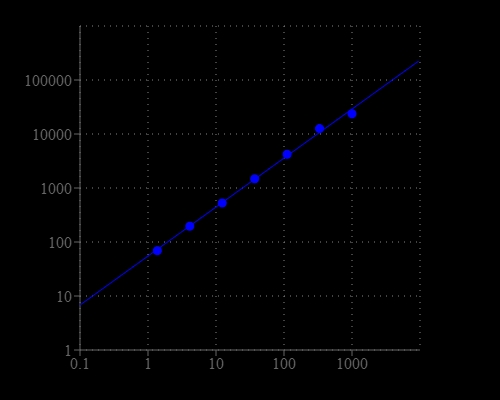
Glutamic acid is one of the 20 proteinogenic amino acids. The carboxylate anions and salts of glutamic acid are known as glutamates. Glutamate is an important neurotransmitter which plays a key role in long-term potentiation and is important for learning and memory. Glutamic acid is the precursor of GABA but has somewhat the opposite function. It might play a role in the normal function of the heart and the prostate. As one of the few nutrients that crosses the blood-brain barrier, glutamic acid is used in the treatment of diseases such as depression, ADD and ADHD, fatigue, alcoholism, epilepsy, muscular dystrophy, mental retardation, and schizophrenia. The Amplite® Fluorimetric Glutamic Acid Assay Kit provides a quick and sensitive method for the measurement of glutamic acid in various biological samples. In the assay, the coupled enzyme system catalyzes the reaction between L-glutamic acid and NADP to produce NADPH, which is specifically recognized by our NADPH sensor and recycled back to NADP. A red flurescence product is produced during the reaction. The signal can be read by either a fluorescence microplate reader or an absorbance microplate reader. With our Amplite® Fluorimetric Glutamic Acid Kit, we have detected as little as 10 µM glutamic acid in a 100 µL reaction volume. The assay is robust, and can be readily adapted for a wide variety of applications that require the measurement of glutamic acid.

Platform


Fluorescence microplate reader

Excitation540 nm
Emission590 nm
Cutoff570 nm
Recommended plateSolid black

Components


Example protocol


AT A GLANCE

Protocol summary

  1. Prepare Glutamic Acid working solution  (50 µL)
  2. Add Glutamic Acid standards and/or test samples (50 µL)
  3. Incubate at room temperature for 30 minutes - 2 hours
  4. Monitor fluorescence intensity at Ex/Em = 540/590 nm (Cutoff = 570 nm)

Important notes
Thaw all the kit components at room temperature before starting the experiment.

PREPARATION OF STOCK SOLUTION

Unless otherwise noted, all unused stock solutions should be divided into single-use aliquots and stored at -20 °C after preparation. Avoid repeated freeze-thaw cycles.

1. NADP stock solution (200X):
Add 100 µL of Dilution Buffer (Component E) into the vial of NADP (Component C) to make 200X NADP stock solution. 

2. Glutamic Acid standard solution (100 mM):
Add 200 µL of Dilution Buffer (Component E) into the vial of Glutamic Acid (Component D) to make 100mM Glutamic Acid standard solution. 


PREPARATION OF STANDARD SOLUTION

Glutamic Acid standard

For convenience, use the Serial Dilution Planner: https://www.aatbio.com/tools/serial-dilution/10054

Take 100 mM Glutamic Acid standard solution and perform 1:100 in Dilution Buffer (Component E) to make 1000 µM Glutamic Acid standard solution (SD7).  Take 1000 µM Glutamic Acid standard solution (SD7) and perform 1:3 serial dilutions to get serially diluted Glutamic Acid standards (SD6 - SD1) with Dilution Buffer (Component E).

PREPARATION OF WORKING SOLUTION

1. Add 10 mL of Assay Buffer (Component B) into the bottle of Enzyme Mix (Component A).

2. Add 50 µL 200X NADP stock solution into the Enzyme Mix bottle, and mix well to make Glutamic Acid working solution.  Note: This Glutamic Acid working solution is enough for two 96-well plates. It is unstable at room temperature, and should be used promptly within 2 hours and avoid exposure to light.  Note: Alternatively, one can make a 50X of Enzyme Mix stock solution by adding 200 μL of H2O into the bottle of Enzyme Mix (Component A), and then prepare the Glutamic Acid working solution by mixing the stock solution with Assay Buffer (Component B) and 200X NADP stock solution proportionally.

SAMPLE EXPERIMENTAL PROTOCOL

Table 1. Layout of Glutamic Acid standards and test samples in a solid black 96-well microplate.  SD = Glutamic Acid Standard, BL = Blank Control, TS = Test Sample. 

BLBLTS TS
SD1SD1......
SD2SD2......
SD3SD3  
SD4SD4  
SD5SD5  
SD6SD6  
SD7 SD7  

Table 2. Reagent composition for each well

WellVolumeReagent
SD1-SD750 µLSerial Dilution (1 to 1000 µM)
BL50 µLDilution Buffer (Component E)
TS50 µLSample
  1. Prepare Glutamic Acid standards (SD), blank controls (BL), and test samples (TS) according to the layout provided in Tables 1 and 2. For a 384-well plate, use 25 µL of reagent per well instead of 50 µL.

  2. Add 50 µL of Glutamic Acid working solution into each well of Glutamic Acid standard, blank control, and test samples to make the total Glutamic Acid assay volume of 100 µL/well. For a 384-well plate, add 25 µL of Glutamic Acid working solution into each well intead, for the total volume of 50 µL/well.

  3. Incubate the reaction at room temperature for 30 minutes to 2 hours, protected from light.

  4. Monitor the fluorescence increase with a fluorescence plate reader at  Excitation = 530 - 570 nm, Emission = 590 - 600 nm (optimal Ex/Em = 540/590 nm), Cutoff = 570 nm.  Note: The contents of the plate can also be transferred to a white clear bottom plate and read by an absorbance microplate reader at the absorbance ratio of ~570 nm to ~605 nm (A575nm/A605nm). The absorption detection has lower sensitivity compared to the fluorescence reading.

Images


Citations


View all 2 citations: Citation Explorer
Experimentally evolved Staphylococcus aureus shows increased survival in the presence of Pseudomonas aeruginosa by acquiring mutations in the amino acid transporter, GltT
Authors: Alexander, Ashley M and Luu, Justin M and Raghuram, Vishnu and Bottacin, Giulia and van Vliet, Simon and Read, Timothy D and Goldberg, Joanna B
Journal: Microbiology (2024): 001445
Presenilin-1/γ-secretase controls glutamate release, tyrosine phosphorylation, and surface expression of N-methyl-d-aspartate receptor (NMDAR) subunit GluN2B
Authors: Xuan, Zhao and Barthet, Gael and Shioi, Junichi and Xu, Jindong and Georgakopoulos, Anastasios and Bruban, Julien and Robakis, Nikolaos K
Journal: Journal of Biological Chemistry (2013): 30495--30501

References


View all 74 references: Citation Explorer
Rational design and synthesis of potent and long-lasting glutamic acid-based dipeptidyl peptidase IV inhibitors
Authors: Tsai TY, Hsu T, Chen CT, Cheng JH, Chiou MC, Huang CH, Tseng YJ, Yeh TK, Huang CY, Yeh KC, Huang YW, Wu SH, Wang MH, Chen X, Chao YS, Jiaang WT.
Journal: Bioorg Med Chem Lett (2009): 1908
Regulation of cerebrospinal fluid levels of cytokines after seizures: the role of IL-6 and glutamic acid
Authors: Lehtimaki KA, Keranen T, Palmio J, Rainesalo S, Saransaari P, Peltola J.
Journal: Eur J Neurol (2009): e75
Glutamic Acid decarboxylase therapy for recent-onset type 1 diabetes: are we at the end or the beginning of finding a cure
Authors: Fleming GA, Klonoff DC.
Journal: J Diabetes Sci Technol (2009): 215
Metabotropic glutamate mGluR5 receptor blockade opposes abnormal involuntary movements and the increases in glutamic acid decarboxylase mRNA levels induced by l-DOPA in striatal neurons of 6-hydroxydopamine-lesioned rats
Authors: Yamamoto N, Soghomonian JJ.
Journal: Neuroscience (2009): 1171
Adsorption of toxic mercury(II) by an extracellular biopolymer poly(gamma-glutamic acid)
Authors: Inbaraj BS, Wang JS, Lu JF, Siao FY, Chen BH.
Journal: Bioresour Technol (2009): 200
Glutamic acid decarboxylase-derived epitopes with specific domains expand CD4(+)CD25(+) regulatory T cells
Authors: Chen G, Han G, Feng J, Wang J, Wang R, Xu R, Shen B, Qian J, Li Y.
Journal: PLoS One (2009): e7034
Binding free energy calculations of N-sulphonyl-glutamic acid inhibitors of MurD ligase
Authors: Perdih A, Bren U, Solmajer T.
Journal: J Mol Model (2009): 983
CE-LIF chiral separation of aspartic acid and glutamic acid enantiomers using human serum albumin and sodium cholate as dual selectors
Authors: Wang S, Fan L, Cui S.
Journal: J Sep Sci (2009): 3184
Isolation and characterization of the promoter sequence of a cassava gene coding for Pt2L4, a glutamic acid-rich protein differentially expressed in storage roots
Authors: de Souza CR, Aragao FJ, Moreira EC, Costa CN, Nascimento SB, Carvalho LJ.
Journal: Genet Mol Res (2009): 334
Effects of incorporation of poly(gamma-glutamic acid) in chitosan/DNA complex nanoparticles on cellular uptake and transfection efficiency
Authors: Peng SF, Yang MJ, Su CJ, Chen HL, Lee PW, Wei MC, Sung HW.
Journal: Biomaterials (2009): 1797