logo
AAT Bioquest

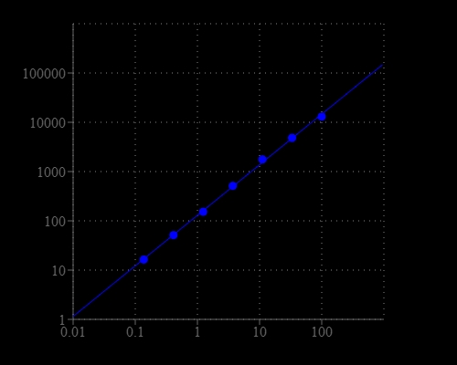
Amplite® Fluorimetric L-Aspartate (Aspartic Acid) Assay Kit

Aspartate dose response was measured with Amplite® Fluorimetric Aspartate Assay Kit on a solid black 96-well plate using a Gemini microplate reader (Molecular Devices).
Ordering information
Price
Catalog Number
Unit Size
Quantity
Add to cart
Additional ordering information
Telephone1-800-990-8053
Fax1-800-609-2943
Emailsales@aatbio.com
InternationalSee distributors
Bulk requestInquire
Custom sizeInquire
ShippingStandard overnight for United States, inquire for international
Request quotation
Storage, safety and handling
H-phraseH303, H313, H333
Hazard symbolXN
Intended useResearch Use Only (RUO)
R-phraseR20, R21, R22
UNSPSC12352200

OverviewpdfSDSpdfProtocol


Aspartate (or Aspartic acid) is a negatively charged, polar amino acid. Aspartate is involved in the control point of pyrimidine biosynthesis, in transamination reactions, interconversions with asparagine, in the metabolic pathway leading to AMP, in the urea cycle, and is a precursor to homoserine, threonine, isoleucine, and methionine. It is also involved in the malate aspartate shuttle. Amplite® Fluorimetric Aspartate Assay Kit offers a sensitive fluorescent assay for quantifying aspartate in biological samples. Aspartate is converted to pyruvate that generates hydrogen peroxide through an enzyme coupled reaction. The amount of hydrogen peroxide generated by aspartate is monitored with Amplite® Red substrate for quantifying aspartate by a fluorescence microplate reader.

Platform


Fluorescence microplate reader

Excitation540 nm
Emission590 nm
Cutoff570 nm
Recommended plateSolid black

Components


Example protocol


AT A GLANCE

Protocol summary

  1. Prepare test samples along with diluted aspartate standards (50 µL)
  2. Add equal volume of working solution (50 µL)
  3. Incubate at 37°C for 20 - 30 minutes
  4. Monitor fluorescence intensity at Ex/Em = 540/590 nm

Important notes
Thaw kit components at room temperature before use. To achieve the best results, it’s recommended to use the black plates.

PREPARATION OF STOCK SOLUTION

Unless otherwise noted, all unused stock solutions should be divided into single-use aliquots and stored at -20 °C after preparation. Avoid repeated freeze-thaw cycles.

1. Aspartate standard solution (10 mM):
Add 100 uL of ddH2O into Aspartate Standard vial (Component E) to make 10 mM aspartate standard solution.

2. Amplite™ Red substrate stock solution (200X):
Add 50 µL of DMSO (Component F) into Amplite™ Red substrate (Component A) to make 200X Amplite™ Red substrate stock solution.

PREPARATION OF STANDARD SOLUTION

Aspartate standard

For convenience, use the Serial Dilution Planner: https://www.aatbio.com/tools/serial-dilution/13827

Add 10 µL of 10 mM aspartate standard into 990 µL of 1X PBS buffer to get 100 µM aspartate solution (ASP7). Then perform 1:3 serial dilutions in 1X PBS buffer to get serially diluted aspartate standards (ASP6 - ASP1).

PREPARATION OF WORKING SOLUTION

1. Conversion Mix solution (100X):
Add 50 µL of ddH2O into Conversion Mix (Component D) to make 100X Conversion Mix stock solution.

2. Amplite™ Red working solution:
Add 5 mL Assay Buffer (Component C) into one Enzyme Mix 1 bottle (Component B1); mix well. Add 100 μL of ddH2O into one Enzyme Mix 2 vial (Component B2); mix well. Transfer the entire vial (100 μL) of Enzyme Mix 2, 25 uL of 200X Amplite™ Red substrate stock solution, and 50 μL of 100X Conversion Mix solution into the Enzyme Mix 1 bottle and mix well. Note: The working solution is not stable, use it promptly, and avoid direct exposure to light.

SAMPLE EXPERIMENTAL PROTOCOL

Table 1. Layout of aspartate standards and test samples in a solid black 96-well microplate. ASP = aspartate standard (ASP1 - ASP7, 0.1 to 100 µM); BL = blank control; TS = test sample.

BLBLTSTS
ASP1ASP1......
ASP2ASP2......
ASP3ASP3  
ASP4ASP4  
ASP5ASP5  
ASP6ASP6  
ASP7ASP7  

Table 2. Reagent composition for each well.

WellVolumeReagent
ASP1 - ASP750 µLSerial Dilution (0.1 to 100 µM)
BL50 µL1X PBS Buffer
TS50 Test Sample
  1. Prepare aspartate standards (ASP), blank controls (BL), and test samples (TS) according to the layout provided in Table 1 and Table 2. For a 384-well plate, use 25 µL of reagent per well instead of 50 µL.

  2. Add 50 µL of Amplite Red™ working solution into each well of aspartate standard, blank control, and test samples to make the total aspartate assay volume of 100 µL/well. For a 384-well plate, add 25 µL of working solution into each well instead, for a total volume of 50 µL/well. Note: Run the aspartate assay at pH 6.5 to 7.0.

  3. Incubate the reaction mixture at 37°C for 20 - 30 minutes.

  4. Monitor the fluorescence increase with a fluorescence plate reader at Ex/Em=540/590 nm (cut off=570 nm).

Images


Citations


View all 1 citations: Citation Explorer
Experimentally evolved Staphylococcus aureus shows increased survival in the presence of Pseudomonas aeruginosa by acquiring mutations in the amino acid transporter, GltT
Authors: Alexander, Ashley M and Luu, Justin M and Raghuram, Vishnu and Bottacin, Giulia and van Vliet, Simon and Read, Timothy D and Goldberg, Joanna B
Journal: Microbiology (2024): 001445

References


View all 20 references: Citation Explorer
A systematic and mechanistic evaluation of aspartic acid as filler for directly compressed tablets containing trimethoprim and trimethoprim aspartate
Authors: ElShaer A, Hanson P, Mohammed AR.
Journal: Eur J Pharm Biopharm (2013): 468
Enzymatic assay for D-aspartic acid using D-aspartate oxidase and oxaloacetate decarboxylase
Authors: Kato S, Ikuta T, Hemmi H, Takahashi S, Kera Y, Yoshimura T.
Journal: Biosci Biotechnol Biochem (2012): 2150
Distribution of free D-aspartic acid and D-aspartate oxidase in frog Rana esculenta tissues
Authors: Di Giovanni M, Burrone L, Chieffi Baccari G, Topo E, Santillo A.
Journal: J Exp Zool A Ecol Genet Physiol (2010): 137
Thyroid hormones and D-aspartic acid, D-aspartate oxidase, D-aspartate racemase, H2O2, and ROS in rats and mice
Authors: Topo E, Fisher G, Sorricelli A, Errico F, Usiello A, D'Aniello A.
Journal: Chem Biodivers (2010): 1467
Aslfm, the D-aspartate ligase responsible for the addition of D-aspartic acid onto the peptidoglycan precursor of Enterococcus faecium
Authors: Bellais S, Arthur M, Dubost L, Hugonnet JE, Gutmann L, van Heijenoort J, Legr and R, Brouard JP, Rice L, Mainardi JL.
Journal: J Biol Chem (2006): 11586
A physiological mechanism to regulate D-aspartic acid and NMDA levels in mammals revealed by D-aspartate oxidase deficient mice
Authors: Errico F, Pirro MT, Affuso A, Spinelli P, De Felice M, D'Aniello A, Di Lauro R.
Journal: Gene (2006): 50
Potassium N-(6-amino-3,4-dihydro-3-methyl-5-nitroso-4-oxopyrimidin-2-yl)-(S)-aspartate N-(6-amino-3,4-dihydro-3-methyl-5-nitroso-4-oxopyrimidin-2-yl)-(S)-aspartic acid 4.88-hydrate: a two-dimensional coordination polymer
Authors: Cuesta R, Glidewell C, Lopez R, Low JN.
Journal: Acta Crystallogr C (2003): m315
Feasibility study: fast liquid chromatography-mass spectrometry for the quantification of aspartic acid in an aspartate drug
Authors: Schmidt J., undefined
Journal: Anal Bioanal Chem (2003): 1120
Modelling the hydrolysis of succinimide: formation of aspartate and reversible isomerization of aspartic acid via succinimide
Authors: Aylin F, Konuklar S, Aviyente V.
Journal: Org Biomol Chem (2003): 2290
Simultaneous analysis of aspartic acid and sorbitol in potassium magnesium aspartate and sorbitol injection by ion-exclusion chromatography with evaporative light-scattering detection
Authors: Wei Y, Ding MY, Zhang WQ.
Journal: Se Pu (2002): 356