Products
Services
Resources
Selection Guides
About
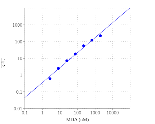
Amplite® Fluorimetric Malondialdehyde (MDA) Quantitation Kit
Enhanced Selectivity
Malondialdehyde (MDA) is one of the natural byproducts during lipid peroxidation. It is widely used as a reliable biomarker to determine oxidative stress, and the quantification of MDA is an essential way to assess oxidative stress in pathophysiological processes. Amplite® Fluorimetric Malondialdehyde (MDA) Quantitation Kit offers a new method to measure MDA without the heating steps required for the TBARS-based MDA assay. The MDA Green™ can react with MDA to generate a considerable enhancement of green fluorescence signal in a convenient 96-well or 384-well microtiter-plate format. Unlike other commercial MDA assay kits, this assay is robust and specific to MDA with little interference from other aldehydes.
| Catalog | Size | Price | Quantity |
|---|---|---|---|
| 10072 | 200 Tests | Price |
Usage and storage
| Intended use | Research Use Only (RUO) |
Instrument settings
| Fluorescence microplate reader | |
| Excitation | 480 nm |
| Emission | 555 nm |
| Cutoff | 530 nm |
| Recommended plate | Solid black |
Contact us
| Telephone | |
| Fax | |
| sales@aatbio.com | |
| International | See distributors |
| Bulk request | Inquire |
| Custom size | Inquire |
| Technical Support | Contact us |
| Request quotation | Request |
| Purchase order | Send to sales@aatbio.com |
| Shipping | Standard overnight for United States, inquire for international |
Page updated on July 17, 2026