logo
AAT Bioquest

Amplite® Fluorimetric Malondialdehyde (MDA) Quantitation Kit

MDA dose response was measured with Amplite® Fluorimetric Malondialdehyde (MDA) Quantitation Kit on a 96-well solid black microplate using a Gemini microplate reader (Molecular Devices) at Ex/Em=365/435 nm, cutoff=420 nm.
Ordering information
Price
Catalog Number
Unit Size
Quantity
Add to cart
Additional ordering information
Telephone1-800-990-8053
Fax1-800-609-2943
Emailsales@aatbio.com
InternationalSee distributors
Bulk requestInquire
Custom sizeInquire
ShippingStandard overnight for United States, inquire for international
Request quotation
Storage, safety and handling
H-phraseH303, H313, H333
Hazard symbolXN
Intended useResearch Use Only (RUO)
R-phraseR20, R21, R22
UNSPSC12171501

OverviewpdfSDSpdfProtocol


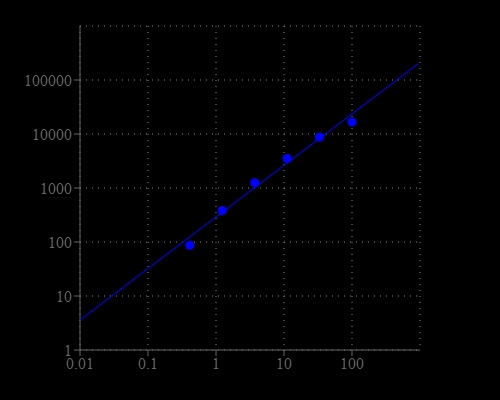
Malondialdehyde (MDA) and 4-hydroxynonenal (4-HNE) are natural byproducts of lipid peroxidation. As the most popular and reliable biomarker for lipid peroxidation, MDA has been widely used for many years to determine oxidative stress in clinical situations. Therefore, quantification of MDA is essential to assess oxidative stress in pathophysiological processes. The Amplite® Fluorimetric Malondialdehyde (MDA) Quantitation Kit offers a quick and convenient method to measure MDA without heating steps that are required for the commercial MDA assay kits from other vendors. Monoaldelite™ Blue itself is nearly non-fluorescent, but generates strong blue fluorescence upon reacting with MDA.

Platform


Fluorescence microplate reader

Excitation365 nm
Emission435 nm
Cutoff420nm
Recommended plateSolid black

Components


Example protocol


AT A GLANCE

Protocol summary

  1. Prepare and add MDA standards or test samples (50 µL)
  2. Prepare and add MDA working solution (50 µL)
  3. Incubate at room temperature for 10 to 30 minutes
  4. Add Reaction Solution (25 µL)
  5. Incubate at room temperature for 30 to 60 minutes
  6. Monitor fluorescence intensity at Ex/Em = 365/435 nm

Important notes
Thaw all the kit components to room temperature before starting the experiment.

PREPARATION OF STOCK SOLUTION

Unless otherwise noted, all unused stock solutions should be divided into single-use aliquots and stored at -20 °C after preparation. Avoid repeated freeze-thaw cycles.

1. MDA standard solution (100 mM):
Add 100 µL of ddH2O into the vial of MDA Standard (Component C) to make 100 mM MDA standard solution.

2. MonoaldeliteTM Blue stock solution (250X):
Add 20 µL of DMSO (Component E) into one vial of MonoaldeliteTM Blue (Component A) to make MonoaldeliteTM Blue stock solution.  Note: This MonoaldeliteTM Blue stock solution is enough for one 96-well plate. It is not stable, use it promptly.

PREPARATION OF STANDARD SOLUTION

MDA standard

For convenience, use the Serial Dilution Planner: https://www.aatbio.com/tools/serial-dilution/10071

Add 10 µL of 100 mM MDA standard solution into 990 µL of Assay Buffer (Component B) to generate 1000 µM MDA standard solution (MDA7). Take 1000 µM MDA standard solution (MDA7) and perform 1:3 serial dilutions to get serially diluted MDA standards (MDA6 - MDA1) with Assay Buffer (Component B).

PREPARATION OF WORKING SOLUTION

MonoaldeliteTM Blue working solution:
Add 20 µL of 250X MonoaldeliteTM Blue stock solution into 5 mL of Assay Buffer (Component B) and mix them well.

SAMPLE EXPERIMENTAL PROTOCOL

Table 1. Layout of MDA standards and test samples in a solid black 96-well microplate. MDA= MDA Standards (MDA1 - MDA7, 1.37 to 1000 µM), BL=Blank Control, TS=Test Samples. 

BLBLTSTS
MDA1MDA1......
MDA2MDA2......
MDA3MDA3  
MDA4MDA4  
MDA5MDA5  
MDA6MDA6  
MDA7MDA7  

Table 2. Reagent composition for each well.

WellVolumeReagent
MDA1 - MDA750 µLSerial Dilutions (1.37 to 1000 µM)
BL50 µLAssay Buffer (Component B)
TS50 µLtest sample
  1. Add MDA standards (MDA), blank controls (BL), and test samples (TS) according to the layout provided in Tables 1 and 2. For a 384-well plate, use 25 µL of reagent per well instead of 50 µL.

  2. Add 50 µL of MDA working solution to each well of MDA standard, blank control, and test samples to make the total assay volume of 100 µL/well. For a 384-well plate, add 25 µL of MDA working solution into each well instead, for a total volume of 50 µL/well.

  3. Incubate the reaction at room temperature for 10 - 30 minutes.

  4. Add 25 µL of Reaction Solution (Component D) to each well to make the total assay volume of 125 µL/well. For a 384-well plate, add 12.5 µL of Reaction Solution (Component D) into each well instead, for a total volume of 62.5 µL/well.

  5. Incubate the reaction at room temperature for 30 - 60 minutes.

  6. Monitor the fluorescence increase with a fluorescence plate reader at Ex/Em = 365/435 nm (Cutoff = 420 nm). 

Images