logo
AAT Bioquest

Amplite® Fluorimetric NADP Assay Kit *Blue Fluorescence*

Nicotinamide adenine dinucleotide (NAD+) and nicotinamide adenine dinucleotide phosphate (NADP+) are two important cofactors for many enzyme reactions found in living cells. NAD forms NADP with the addition of a phosphate group to the 2' position of the adenyl nucleotide through an ester linkage. NADP is used in anabolic biological reactions, such as fatty acid and nucleic acid synthesis, which requires NADPH as a reducing agent. In chloroplasts, NADP is an oxidizing agent important in the preliminary reactions of photosynthesis. The NADPH produced by photosynthesis is used as reducing power for the biosynthetic reactions in the Calvin cycle of photosynthesis. Quantifying the generation or consumption of these factors is an important method to monitor the enzyme-mediated reaction or screening the modulator or substrate of these enzyme reactions. There are several kits on the market to quantify NADPH or total NADP/NADPH amount, but detection NADP generation in the presence of large excess amount of NADPH has been quite challenging to date because NADP has its absorption peak at 260 nm and does not fluorescence, making the measurement unpractical. Amplite® Fluorimetric NADP Assay Kit provides a sensitive and rapid detection of NADP. The kit directly measure NADP using Quest Fluor™ NADP reagent, our newly developed NADP sensor. The proprietary probe used in this kit reacts only with NADP to generate a product that fluorescence at a specific excitation and emission spectra range and has little response to NADPH. This kit can detect as little as 30 nM NADP in a 100 µL assay volume, and monitor 0.3% NADP generation in the presence of excess amount of NADPH. This assay can be performed in a convenient 96-well or 384-well microtiter-plate format and can be used in high-throughput screening.
Example protocol

AT A GLANCE

Protocol Summary
  1. Prepare NADP standards or test samples (50 µL)
  2. Add 20 µL Quest Fluor™ NADP Probe
  3. Add 20 µL Assay Solution
  4. Incubate at RT for 10 - 20 minutes
  5. Add 15 µL Enhancer Solution
  6. Incubate at RT for 10 - 20 minutes
  7. Monitor Fluorescence at 420/480 nm (Cutoff = 455 nm)
Important Note

Thaw each kit components at room temperature before starting the experiment.

PREPARATION OF STOCK SOLUTIONS

Unless otherwise noted, all unused stock solutions should be divided into single-use aliquots and stored at -20 °C after preparation. Avoid repeated freeze-thaw cycles

NADP standard solution (1 mM)

Add 500 µL of ddH2O into the vial of NADP Standard (Component D) to make 1 mM NADP standard solution.

PREPARATION OF STANDARD SOLUTIONS

For convenience, use the Serial Dilution Planner:
https://www.aatbio.com/tools/serial-dilution/15281

NADP standard
Add 10 µL of 1mM NADP standard solution into 990 µL ddH2O or 1X PBS buffer to generate 10 µM NADP standard solution (NS7). Take 10 µM NADP standard solution (NS7) and perform 1:3 serial dilutions in ddH2O or 1X PBS buffer to get serially diluted NADP standard (NS6 - NS1). Note: Diluted NADP standard solution is unstable, and should be used within 4 hours.

SAMPLE EXPERIMENTAL PROTOCOL

Table 1. Layout of NADP standards and test samples in a black/solid bottom 96-well microplate. NS=NADP standards (NS1-NS7, 0.01 to 10 µM); BL= blank control; TS=test sample.

BLBLTSTS
NS1NS1......
NS2NS2......
NS3NS3
NS4NS4
NS5NS5
NS6NS6
NS7NS7

Table 2. Reagent composition for each well.

WellVolumeReagent
NS1-NS750 µLserial dilution (0.01 to 10 µM)
BL50 µL

ddH2O or 1X PBS buffer

TS50 µLsample
  1. Prepare NADP standards (NS), blank controls (BL), and test samples (TS) according to the layout provided in Table 1 and Table 2. For a 384-well plate, use 25 µL of reagent per well instead of 50 µL.
  2. Add 20 µL Quest Fluor™ NADP Probe (Component A) solution into each well of NADP standard, blank control, and test samples, mix well. For a 384-well plate, use 10 µL of Quest Fluor™ NADP Probe (Component A) solution instead.
  3. Add 20 µL Assay Solution (Component B) into each well, mix well. For a 384-well plate, use 10 µL of Assay Solution (Component B) instead.
  4. Incubate the reaction at room temperature for 10 - 20 minutes, protected from light.
  5. Add 15 µL Enhancer (Component C) to each well to make the total NADP assay volume of 105 µL/well. For a 384-well plate, add 7.5 uL Enhancer (Component C) instead, for a total volume of 52.5 µL/well.
  6. Incubate at room temperature for 10 - 20 minutes, protected from light.
  7. Monitor the fluorescence increase with a fluorescence plate reader at 420/480 nm (Cutoff = 455nm).

Citations

View all 57 citations: Citation Explorer
Resveratrol attenuates excessive ethanol exposure induced insulin resistance in rats via improving NAD+/NADH ratio
Authors: Luo, Gang and Huang, Bingqing and Qiu, Xiang and Xiao, Lin and Wang, Ning and Gao, Qin and Yang, Wei and Hao, Liping
Journal: Molecular Nutrition & Food Research (2017)
Epigenetic regulation of Runx2 transcription and osteoblast differentiation by nicotinamide phosphoribosyltransferase
Authors: Ling, Min and Huang, Peixin and Islam, Shamima and Heruth, Daniel P and Li, Xuanan and Zhang, Li Qin and Li, Ding-You and Hu, Zhaohui and Ye, Shui Qing
Journal: Cell & Bioscience (2017): 27
MCU-dependent mitochondrial Ca2+ inhibits NAD+/SIRT3/SOD2 pathway to promote ROS production and metastasis of HCC cells
Authors: Ren, T and Zhang, H and Wang, J and Zhu, J and Jin, M and Wu, Y and Guo, X and Ji, L and Huang, Q and Yang, H and others, undefined
Journal: Oncogene (2017)
Metabolic and molecular insights into an essential role of nicotinamide phosphoribosyltransferase
Authors: Zhang, Li Q and Van Ha, undefined and el, Leon and Xiong, Min and Huang, Peixin and Heruth, Daniel P and Bi, Charlie and Gaedigk, Roger and Jiang, Xun and Li, Ding-You and Wyckoff, Gerald and others, undefined
Journal: Cell Death & Disease (2017): e2705
Cytosolic Redox Status of Wine Yeast (Saccharomyces Cerevisiae) under Hyperosmotic Stress during Icewine Fermentation
Authors: Yang, Fei and Heit, Caitlin and Inglis, Debra L
Journal: Fermentation (2017): 61

References

View all 1 references: Citation Explorer
Inhibition of leucine aminopeptidase 3 suppresses invasion of ovarian cancer cells through down-regulation of fascin and MMP-2/9
Authors: Wang X, Shi L, Deng Y, Qu M, Mao S, Xu L, Xu W, Fang C.
Journal: Eur J Pharmacol (2015): 116
Page updated on December 6, 2024

Ordering information

Price
Unit size
Catalog Number15281
Quantity
Add to cart

Additional ordering information

Telephone1-800-990-8053
Fax1-800-609-2943
Emailsales@aatbio.com
InternationalSee distributors
Bulk requestInquire
Custom sizeInquire
Technical SupportContact us
Purchase orderSend to sales@aatbio.com
ShippingStandard overnight for United States, inquire for international
Request quotation

Storage, safety and handling

H-phraseH303, H313, H333
Hazard symbolXN
Intended useResearch Use Only (RUO)
R-phraseR20, R21, R22
UNSPSC12352200

Platform

Fluorescence microplate reader

Excitation420 nm
Emission480 nm
Cutoff455 nm
Recommended plateSolid black

Components

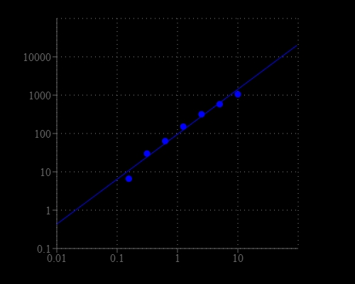
NADP standard curve with 100 µM NADPH in presence in the solution. As low as 0.3% of NADP (~300 nM) converted from NADPH can be detected with 20 min incubation (n=3). RFU read at Ex/Em = 420/480 nm.
Comparison of NADP and NADPH response