logo
AAT Bioquest

Amplite® Fluorimetric Proteasome 20S Activity Assay Kit *Green Fluorescence*

Detection of proteasome activity in Jurkat cells with Amplite® Fluorimetric Proteasome 20S Activity Assay Kit. Jurkat cells were seeded on the same day at 500,000 cells/90 &micro;L/well in a 96-well black wall/clear bottom Costar plate. The cells were treated with or without 50 mM H<sub>2</sub>O<sub>2</sub> for 30 minutes. The proteasome assay loading solution (100 &micro;L/well) was added and incubated in a 5% CO2, 37 &deg;C incubator for 3 hours. The fluorescence intensity was measured at Ex/Em = 490/525 by using a Gemini fluorescent microplate reader (Molecular Devices).
Ordering information
Price
Catalog Number
Unit Size
Quantity
Add to cart
Additional ordering information
Telephone1-800-990-8053
Fax1-800-609-2943
Emailsales@aatbio.com
InternationalSee distributors
Bulk requestInquire
Custom sizeInquire
ShippingStandard overnight for United States, inquire for international
Request quotation
Spectral properties
Extinction coefficient (cm -1 M -1)80000
Excitation (nm)500
Emission (nm)522
Storage, safety and handling
H-phraseH303, H313, H333
Hazard symbolXN
Intended useResearch Use Only (RUO)
R-phraseR20, R21, R22
UNSPSC12352200

OverviewpdfSDSpdfProtocol


Extinction coefficient (cm -1 M -1)
80000
Excitation (nm)
500
Emission (nm)
522
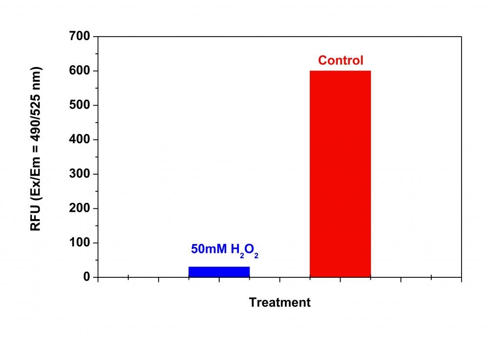
Our Amplite® Fluorometric 20S Proteasome Assay Kit is a homogeneous fluorescent assay that measures the chymotrypsin-like protease activity associated with the proteasome complex either in cultured cells or cell lysates. This kit uses LLVY-R110 as a fluorogenic indicator for proteasome activity. Cleavage of LLVY-R110 by proteasome generates the strongly green fluorescent R110 that is monitored fluorimetrically at 520-530 nm with excitation of 480-500 nm. The kit provides all the essential components with an optimized assay protocol. The assay is robust, and can be readily adapted for high-throughput assays for evaluation of proteasome activity or inhibitor screening in cultured cells or in solution. The assay can be performed in a convenient 96-well and 384-well fluorescence microtiter-plate format. The main function of the proteasome is to degrade unneeded or damaged proteins by proteolysis, a chemical reaction that breaks peptide bonds. The proteasomal degradation pathway is essential for many cellular processes, including the cell cycle, the regulation of gene expression, and responses to oxidative stress. The most common form of the proteasome in this pathway is the 26S proteasome, an ATP-dependent proteolytic complex, which contains one 20S (700-kDa) core particle structure and two 19S (700-kDa) regulatory caps. The 20S core contains three major proteolytic activities including chymotrypsin-like, trypsin-like and caspase-like. It is responsible for the breakdown of key proteins involved with apoptosis, DNA repair, endocytosis, and cell cycle control.

Platform


Fluorescence microplate reader

Excitation490 nm
Emission525 nm
Cutoff515 nm
Recommended plateSolid black
Instrument specification(s)Top read mode

Components


Example protocol


AT A GLANCE

Protocol Summary
  1. Prepare cells with test compounds (100 µL/well/96-well plate or 25 µL/well/384-well plate)
  2. Add equal volume of Proteasome working solution (100 µL/well/96-well plate or 25 µL/well/384-well plate)
  3. Incubate at 37°C for at least 1 hour

  4. Monitor fluorescence intensity at Ex/Em = 490/525 nm (Cutoff = 515 nm)
Important Note

Thaw all the kit components at room temperature before use.

CELL PREPARATION

For guidelines on cell sample preparation, please visit https://www.aatbio.com/resources/guides/cell-sample-preparation.html

PREPARATION OF STOCK SOLUTIONS

Unless otherwise noted, all unused stock solutions should be divided into single-use aliquots and stored at -20 °C after preparation. Avoid repeated freeze-thaw cycles

Proteasome LLVY-R110 Substrate stock solution (400X)

Add 25 µL of DMSO (Component C) to the vial of Proteasome LLVY-R110 Substrate (Component A), and mix well to make 400X Proteasome LLVY-R110 Substrate stock solution.

PREPARATION OF WORKING SOLUTION

Add 25 μL of 400X Proteasome LLVY-R110 Substrate stock solution into 10 mL of Assay Buffer (Component B) and mix well to make Proteasome working solution. Note: This Proteasome working solution is enough for 1 plate. Protect from light.

SAMPLE EXPERIMENTAL PROTOCOL

  1. Treat cells with 10 µL of 10X test compound (for a 96-well plate) or 5 µL of 5X test compound (for a 384-well plate) in PBS or desired buffer. For blank wells (medium without the cells), add the corresponding amount of compound buffer.
  2. Incubate the cell plates in a 5% CO2, 37°C incubator for a desired period of time. Note: Pure proteasome or cell lysates can be used directly for screening the proteasome inhibitors.

  3. Add 100 µL/well (96-well plate) or 25 µL/well (384-well plate) of Proteasome working solution.
  4. Incubate the plate at 37°C for at least 1 hour (2 hours to overnight), protected from light. Note: Each cell line should be evaluated on an individual basis to determine the optimal incubation time.

  5. Monitor the fluorescence intensity (top read) at Ex/Em = 490/525 nm (Cutoff = 515 nm).

Spectrum


Open in Advanced Spectrum Viewer
spectrum

Spectral properties

Extinction coefficient (cm -1 M -1)80000
Excitation (nm)500
Emission (nm)522

Images


Citations


View all 13 citations: Citation Explorer
Quantitative reactive cysteinome profiling reveals a functional link between ferroptosis and proteasome-mediated degradation
Authors: Wang, Yankun and Wang, Chu
Journal: Cell Death \& Differentiation (2022): 1--12
TRIB2 modulates proteasome function to reduce ubiquitin stability and protect liver cancer cells against oxidative stress
Authors: Guo, Susu and Chen, Yuxin and Yang, Yueyue and Zhang, Xiao and Ma, Lifang and Xue, Xiangfei and Qiao, Yongxia and Wang, Jiayi
Journal: Cell death \& disease (2021): 1--18
TRIB2 desensitizes ferroptosis via $\beta$TrCP-mediated TFRC ubiquitiantion in liver cancer cells
Authors: Guo, Susu and Chen, Yuxin and Xue, Xiangfei and Yang, Yueyue and Wang, Yikun and Qiu, Shiyu and Cui, Jiangtao and Zhang, Xiao and Ma, Lifang and Qiao, Yongxia and others,
Journal: Cell death discovery (2021): 1--13
MnFe2O4 nanoparticles accelerate the clearance of mutant huntingtin selectively through ubiquitin-proteasome system
Authors: Zhang, Li and Wei, Peng-Fei and Song, Yong-Hong and Dong, Liang and Wu, Ya-Dong and Hao, Zong-Yao and Fan, Song and Tai, Sheng and Meng, Jia-Lin and Lu, Yang and others, undefined
Journal: Biomaterials (2019): 119248
Melatonin Promotes Ubiquitination of Phosphorylated Pro-Apoptotic Protein Bcl-2-Interacting Mediator of Cell Death-Extra Long (BimEL) in Porcine Granulosa Cells
Authors: Wang, Yingzheng and Zeng, Shenming
Journal: International journal of molecular sciences (2018): 3431
Photothermally Controlled MHC Class I Restricted CD8+ T-Cell Responses Elicited by Hyaluronic Acid Decorated Gold Nanoparticles as a Vaccine for Cancer Immunotherapy
Authors: Cao, Fengqiang and Yan, Mengmeng and Liu, Yijia and Liu, Lanxia and Ma, Guilei
Journal: Advanced healthcare materials (2018): 1701439
Mycobacterium tuberculosis controls phagosomal acidification by targeting CISH-mediated signaling
Authors: Queval, Christophe J and Song, Ok-Ryul and Carralot, Jean-Philippe and Saliou, Jean-Michel and Bongiovanni, Antonino and Deloison, Gaspard and Deboosere, Nathalie and Jouny, Samuel and Iantomasi, Raffaella and Delorme, Vincent and others,
Journal: Cell reports (2017): 3188--3198
Interactome analysis reveals a novel role for RAD6 in the regulation of proteasome activity and localization in response to DNA damage
Authors: An, Hongli and Yang, Lu and Wang, Chen and Gan, Zhixue and Gu, Haihui and Zhang, Tao and Huang, Xin and Liu, Yan and Li, Yufeng and Chang, Shing-Jyh and others,
Journal: Molecular and cellular biology (2017): e00419--16
Mycobacterium tuberculosis Controls Phagosomal Acidification by Targeting CISH-Mediated Signaling
Authors: Queval, Christophe J and Song, Ok-Ryul and Carralot, Jean-Philippe and Saliou, Jean-Michel and Bongiovanni, Antonino and Deloison, Gaspard and Deboos&egrave;re, Nathalie and Jouny, Samuel and Iantomasi, Raffaella and Delorme, Vincent and others, undefined
Journal: Cell Reports (2017): 3188--3198
Autophagy-mediated clearance of ubiquitinated mutant huntingtin by graphene oxide
Authors: Jin, Peipei and Wei, Pengfei and Zhang, Yunjiao and Lin, Jun and Sha, Rui and Hu, Yi and Zhang, Jiqian and Zhou, Wei and Yao, Han and Ren, Li and others, undefined
Journal: Nanoscale (2016)