logo
AAT Bioquest

Amplite® Human Serum Albumin (HSA) Site I Binding Assay Kit

Response of Warfarin (Site-1 drug) and Ibuprofen (Site-2 drug) was measured using Amplite® Human Serum Albumin (HSA) Site I Binding Assay Kit. The response was acquired using Spectramax Gemini XS (Molecular devices) with Ex/Em = 365/480 nm with cutoff = 435 nm.
Ordering information
Price
Catalog Number
Unit Size
Quantity
Add to cart
Additional ordering information
Telephone1-800-990-8053
Fax1-800-609-2943
Emailsales@aatbio.com
InternationalSee distributors
Bulk requestInquire
Custom sizeInquire
ShippingStandard overnight for United States, inquire for international
Request quotation
Spectral properties
Excitation (nm)334
Emission (nm)511
Storage, safety and handling
H-phraseH303, H313, H333
Hazard symbolXN
Intended useResearch Use Only (RUO)
R-phraseR20, R21, R22
UNSPSC12171501

OverviewpdfSDSpdfProtocol


Excitation (nm)
334
Emission (nm)
511
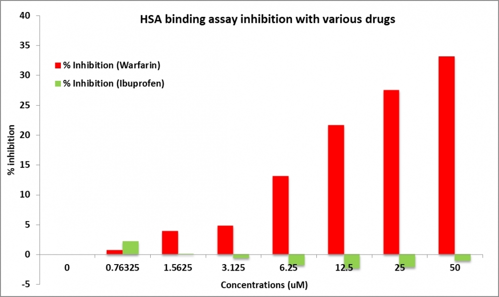
Human serum albumin (HSA) is one of the most important carriers for acidic drugs in human plasma and has been shown to bind a large number of different compounds in a reversible manner. Several different ligand binding sites have been identified for HSA. Among them, Site I has been identified as one of major drug binding sites. Amplite®™ Human Serum Albumin (HSA) Site I Binding Assay Kit is a fluorescence-based high throughput assay to determine the small molecule binding towards HSA. This assay is based on a novel fluorescent probe, HSA Blue™ S1. It has been characterized to bind to the site 1 of HSA with unique spectroscopic and binding properties. HSA Blue™ S1 displays a large fluorescence intensity difference between the protein-bound and protein-unbound state. The competition of small molecules for HSA binding in the presence of HSA Blue™ S1 results in low fluorescence intensities. This assay can be used as a high throughput screen tool to determine total binding to HSA at Site I.

Platform


Fluorescence microplate reader

Excitation365 nm
Emission480 nm
Cutoff435 nm
Recommended plateSolid black
Instrument specification(s)Top read mode

Components


Example protocol


AT A GLANCE

Protocol Summary
  1. Add HSA working solution (50 µL) and HSA Blue™ S1 working solution (50 µL) to the wells
  2. Add test drugs (50 µL) with various concentrations to respective wells
  3. Incubate for 15 to 45 minutes at RT
  4. Measure response with fluorescence microplate reader at Ex/Em = 365/480 nm (Cutoff = 435 nm) 

Important
Bring all the kit components at room temperature before starting the experiment.

PREPARATION OF STOCK SOLUTIONS

Unless otherwise noted, all unused stock solutions should be divided into single-use aliquots and stored at -20 °C after preparation. Avoid repeated freeze-thaw cycles.

HSA Blue™ S1 stock solution
Add 100 µL DMSO (Component D) into HSA Blue™ S1 (Component A) and mix well.
Note     Store the unused HSA Blue™ S1 stock solution at -20 °C in single use aliquots to avoid freeze thaw cycles.

PREPARATION OF WORKING SOLUTION

1. HSA Blue™ S1 working solution
Add 50 µL HSA Blue™ S1 stock solution into 5 mL of HSA Assay Buffer (Component B) and mix well.
Note     HSA Blue™ S1 working solution should not be stored and should be used promptly.
Note     5 mL HSA Blue™ S1 working solution is enough for one 96-well plate.


2. HSA working solution
Add 250 µL HSA solution (Component C) into 5 mL of HSA Assay Buffer (Component B) and mix well.
Note     5 mL HSA working solution is enough for one 96-well plate.


3. Test drugs working solution
Dilute drugs stock solution to the desired concentrations in 3X working solutions using HSA Assay Buffer (Component B).
Note     For the protocol mentioned here, suggested volume for the one well is 50 µL.

SAMPLE EXPERIMENTAL PROTOCOL

The following protocol can be used as a guideline and should be optimized according to the needs.
  1. Add 50 µL of HSA Blue™ S1 working solution in wells.
  2. Add 50 µL of HSA working solution in wells.
  3. Add 50 µL of drugs working solution to their respective wells. (Total volume = 150 µL/well).
  4. Incubate the samples for 30 minutes at RT.
  5. Monitor the fluorescence increase with a fluorescence plate reader at Ex/Em = 365/480 nm (Cutoff = 435 nm). 

Spectrum


Open in Advanced Spectrum Viewer
spectrum

Spectral properties

Excitation (nm)334
Emission (nm)511

Product Family


Images


References


View all 50 references: Citation Explorer
Anionic versus neutral Pt(II) complexes: The relevance of the charge for human serum albumin binding.
Authors: Ricciardi, Loredana and Guzzi, Rita and Rizzuti, Bruno and Ionescu, Andreea and Aiello, Iolinda and Ghedini, Mauro and La Deda, Massimo
Journal: Journal of inorganic biochemistry (2020): 111024
Human Serum Albumin Binding in a Vial: A Novel UV-pH Titration Method To Assist Drug Design.
Authors: Dargó, Gergő and Bajusz, Dávid and Simon, Kristóf and Müller, Judit and Balogh, György T
Journal: Journal of medicinal chemistry (2020): 1763-1774
Anticancer activity, calf thymus DNA and human serum albumin binding properties of Farnesiferol C from Ferula pseudalliacea.
Authors: Tanzadehpanah, Hamid and Mahaki, Hanie and Samadi, Pouria and Karimi, Jamshid and Moghadam, Neda Hosseinpour and Salehzadeh, Sadegh and Dastan, Dara and Saidijam, Massoud
Journal: Journal of biomolecular structure & dynamics (2019): 2789-2800
An integrated quantitative structure and mechanism of action-activity relationship model of human serum albumin binding.
Authors: Serra, Angela and Önlü, Serli and Coretto, Pietro and Greco, Dario
Journal: Journal of cheminformatics (2019): 38
The displacement study of 99m Tc-DTPA-Human serum albumin binding in presence of furosemide and metformin by using equilibrium dialysis and molecular docking.
Authors: Chemlal, Laila and Akachar, Jihane and Makram, Sanaa and Zoubir, Brahim and Cherrah, Yahia and Eljaoudi, Rachid and Ibrahimi, Azeddine and Faouzi, Mly A
Journal: IUBMB life (2019): 2003-2009
Redox properties and human serum albumin binding of nitro-oleic acid.
Authors: Zatloukalova, Martina and Mojovic, Milos and Pavicevic, Aleksandra and Kabelac, Martin and Freeman, Bruce A and Pekarova, Michaela and Vacek, Jan
Journal: Redox biology (2019): 101213
Multi-Spectroscopic Characterization of Human Serum Albumin Binding with Cyclobenzaprine Hydrochloride: Insights from Biophysical and In Silico Approaches.
Authors: Baig, Mohammad Hassan and Rahman, Safikur and Rabbani, Gulam and Imran, Mohd and Ahmad, Khurshid and Choi, Inho
Journal: International journal of molecular sciences (2019)
Comparative studies on the human serum albumin binding of the clinically approved EGFR inhibitors gefitinib, erlotinib, afatinib, osimertinib and the investigational inhibitor KP2187.
Authors: Dömötör, Orsolya and Pelivan, Karla and Borics, Attila and Keppler, Bernhard K and Kowol, Christian R and Enyedy, Éva A
Journal: Journal of pharmaceutical and biomedical analysis (2018): 321-331
Human serum albumin binding to the biologically active labdane diterpene "leoheterin": Spectroscopic and in silico analysis.
Authors: Ali, Mohd Sajid and Amina, Musarat and Al-Lohedan, Hamad A and Al Musayeib, Nawal M
Journal: Journal of photochemistry and photobiology. B, Biology (2018): 9-17
Alteration of human serum albumin binding properties induced by modifications: A review.
Authors: Maciążek-Jurczyk, Małgorzata and Szkudlarek, Agnieszka and Chudzik, Mariola and Pożycka, Jadwiga and Sułkowska, Anna
Journal: Spectrochimica acta. Part A, Molecular and biomolecular spectroscopy (2018): 675-683