logo
AAT Bioquest

Amplite® Luciferase Reporter Gene Assay Kit *Maximized Luminescence*

Reaction Kinetics of CHO-V<sub>2</sub>R-Luc cells using Amplite® Luciferase Reporter Gene Assay Kit. CHO cells stably transfected with pCRE-luciferase gene and human Vasopressin receptor 2 (V<sub>2</sub>R) were plated into a 384-well white wall/clear bottom costar plate at 15,000 cells/well/25 &micro;L. Cells then were treated with 100 nM of vasopressin in a 5% CO<sub>2</sub> incubator at 37 &deg;C for 4 hours. 25 &micro;L of luciferase assay solution was added into the well. The kinetic data was taken every 30 minutes for up to 3 hours with a NOVOstar plate reader (BMG Labtech). The vasopressin induced luciferase signal is stable for more than 3 hours.
Ordering information
Price
Catalog Number
Unit Size
Quantity
Add to cart
Additional ordering information
Telephone1-800-990-8053
Fax1-800-609-2943
Emailsales@aatbio.com
InternationalSee distributors
Bulk requestInquire
Custom sizeInquire
ShippingStandard overnight for United States, inquire for international
Request quotation
Storage, safety and handling
H-phraseH303, H313, H333
Hazard symbolXN
Intended useResearch Use Only (RUO)
R-phraseR20, R21, R22
UNSPSC41105331

OverviewpdfSDSpdfProtocol


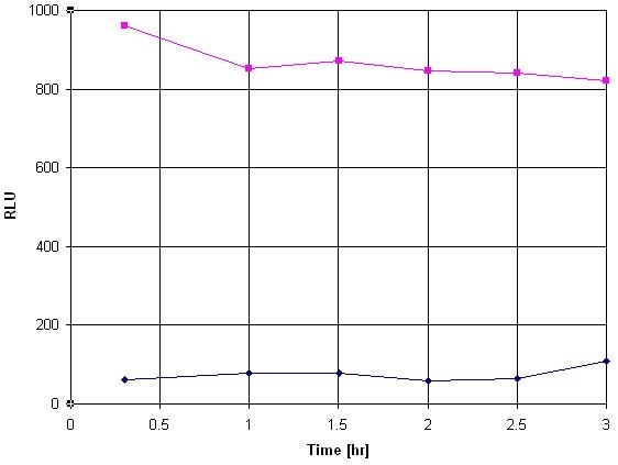
Common reporter genes include beta-galactosidase, beta-glucuronidase and luciferase. The advantages of a luciferase assay are the high sensitivity, the absence of luciferase activity inside most of the cell types, the wide dynamic range, rapidity and low cost. The most versatile and common reporter gene is the luciferase of the North American firefly Photinus pyralis. The protein requires no posttranslational modification for enzyme activity. It is not even toxic in high concentration (in vivo) and can be used in pro- and eukaryotic cells. The firefly luciferase catalyzes the bioluminescent oxidation of luciferin in the presence of ATP, magnesium and oxygen. This Amplite® Luciferase Reporter Gene Assay Kit uses a proprietary luminogenic formulation to quantify luciferase activity in live cells and cell extracts. Our formulation generates a luminescent product that gives strong luminescence upon interaction with luciferase. The kit provides all the essential components with our optimized 'mix and read' assay protocol that is compatible with HTS liquid handling instruments. It has extremely high sensitivity, and can be used for the assays that require demanding sensitivity.

Platform


Luminescence microplate reader

Recommended plateSolid white

Components


Example protocol


AT A GLANCE

Protocol summary

  1. Prepare cells (samples) with test compounds (100 µL/well for 96-well plate or 25 µL/well for 384-well plate)
  2. Add equal volume of Luciferase Sensor working solution
  3. Incubate at room temperature for 10 - 20 minutes
  4. Monitor luminescence intensity at 560 nm

Important notes
Thaw all the kit components to room temperature before use. For all luminescent experiments, it is recommended to use white plates to get the best results.

PREPARATION OF WORKING SOLUTION

1. For Cat.# 12518, transfer the whole content of Reaction Buffer (Component B) into the bottle of Luciferase Sensor (Component A) and mix well to make Luciferase Sensor working solution. 

2. For Cat.# 12519, add 10 mL of Reaction Buffer (Component B) and for Cat.# 12520, add 100 mL of Reaction Buffer (Component B) into the bottle of Luciferase Sensor (Component A) and mix well.  Then, transfer the resulted solution back to the bottle of Reaction Buffer (Component B).  Multiple washes are necessary to completely transfer the contents. Note: The reconstituted Luciferase Sensor working solution is not stable. Protect from light.

For guidelines on cell sample preparation, please visit
https://www.aatbio.com/resources/guides/cell-sample-preparation.html

SAMPLE EXPERIMENTAL PROTOCOL

Run Luciferase assay:

  1. Treat cells (or samples) with test compounds by adding 10 µL of 10X test compounds (96-well plate) or 5 µL of 5X test compounds (384-well plate) in desired compound buffer. For blank wells (medium without the cells), add the corresponding amount of compound buffer.

  2. Incubate the cell plate in a 5% CO2 incubator at 37°C for desired period of time, typically 4 hours to overnight.

  3. Add 100 µL (96-well plate) or 25 µL (384-well plate) per well of Luciferase Sensor working solution.

  4. Incubate the plate at room temperature for 10 - 20 minutes. Keep from light.

  5. Monitor luminescence intensity with a luminometer.

Establish standard Luciferase calibration curve: Note: Luciferase standard curve should be generated together with the above assay if the absolute amount of Luciferase in samples needs to be calculated.

  1. Make a series dilutions of Luciferase in PBS buffer with 0.1% BSA by including a sample without Luciferase (as a control) for measuring background luminescence. Note: Typically Luciferase concentrations from 1 pg/mL to 1 ng/mL are appropriate.

  2. Add 100 µL/well (96-well plate) or 25 µL/well (384-well plate) of diluted Luciferase solution into an empty plate.

  3. Add 100 µL/well (96-well plate) or 25 µL/well (384-well plate) of Luciferase working solution.

  4. Incubate the reaction mixture at room temperature for 10 - 20 minutes, protected from light.

  5. Record the luminescence intensity with a standard luminometer.

  6. Generate the Luciferase standard curve.

Images


Citations


View all 10 citations: Citation Explorer
SARS-CoV-2 omicron variants harbor spike protein mutations responsible for their attenuated fusogenic phenotype
Authors: Park, Seung Bum and Khan, Mohsin and Chiliveri, Sai Chaitanya and Hu, Xin and Irvin, Parker and Leek, Madeleine and Grieshaber, Ailis and Hu, Zongyi and Jang, Eun Sun and Bax, Ad and others,
Journal: Communications Biology (2023): 556
Discovery of small molecule agonists of the Relaxin Family Peptide Receptor 2
Authors: Esteban-Lopez, Maria and Wilson, Kenneth J and Myhr, Courtney and Kaftanovskaya, Elena M and Henderson, Mark J and Southall, Noel T and Xu, Xin and Wang, Amy and Hu, Xin and Barnaeva, Elena and others,
Journal: Communications biology (2022): 1--12
AtHSPR is involved in GA-and light intensity-mediated control of flowering time and seed set in Arabidopsis
Authors: Yang, Tao and Sun, Yan and Wang, Yongli and Zhou, Lina and Chen, Mengya and Bian, Zhiyuan and Lian, Yuke and Xuan, Lijuan and Yuan, Guoqiang and Wang, Xinyu and others,
Journal: Journal of Experimental Botany (2020)
AtHSPR is involved in GA-and light intensity-mediated control of flowering time and seed set in Arabidopsis
Authors: Yang, Tao and Sun, Yan and Wang, Yongli and Zhou, Lina and Chen, Mengya and Bian, Zhiyuan and Lian, Yuke and Xuan, Lijuan and Yuan, Guoqiang and Wang, Xinyu and others,
Journal: Journal of experimental botany (2020): 3543--3559
LncRNA TUBA4B functions as a competitive endogenous RNA to inhibit gastric cancer progression by elevating PTEN via sponging miR-214 and miR-216a/b
Authors: Guo, Jianbo and Li, Yan and Duan, He and Yuan, Lu
Journal: Cancer Cell International (2019): 156
Microwave ablation-assisted liver gene transfection in rats
Authors: Jiang, Ruoyu and Meng, Lingkai and Sun, Longhao and He, Xianghui and Liang, Xiaoyu and Zhang, Jie and Zhang, Zhixiang
Journal: International Journal of Hyperthermia (2016): 666--672
Identification of compounds that modulate retinol signaling using a cell-based qHTS assay
Authors: Chen, Yanling and Sakamuru, Srilatha and Huang, Ruili and Reese, David H and Xia, Menghang
Journal: Toxicology in Vitro (2016): 287--296
Activation of relaxin family receptor 1 from different mammalian species by relaxin peptide and small-molecule agonist ML290
Authors: Huang, Zaohua and Myhr, Courtney and Bathgate, Ross AD and Ho, Brian A and Bueno, Amaya and Hu, Xin and Xiao, Jingbo and Southall, Noel and Barnaeva, Elena and Agoulnik, Irina U and others, undefined
Journal: Frontiers in endocrinology (2015)
Neuroprotective effect of schizandrin A on oxygen and glucose deprivation/reperfusion-induced cell injury in primary culture of rat cortical neurons
Authors: Wang, Cai-Ping and Li, Gui-Cai and Shi, Yun-Wei and Zhang, Xiao-Chuan and Li, Jian-Long and Wang, Zhi-Wei and Ding, Fei and Liang, Xin-Miao
Journal: Journal of physiology and biochemistry (2014): 735--747
Discovery of ML367, inhibitor of ATAD5 stabilization
Authors: Rohde, Jason M and Rai, Ganesha and Choi, Yong Jun and Sakamuru, Srilatha and Fox, Jennifer T and Huang, Ruili and Xia, Menghang and Myung, Kyungjae and Boxer, Matthew B and Maloney, David J
Journal: (2013)

References


View all 52 references: Citation Explorer
In vivo bioluminescence tumor imaging of RGD peptide-modified adenoviral vector encoding firefly luciferase reporter gene
Authors: Niu G, Xiong Z, Cheng Z, Cai W, Gambhir SS, Xing L, Chen X.
Journal: Mol Imaging Biol (2007): 126
Creation and Characterization of a Doxycycline-Inducible Mouse Model of Thyroid-Targeted RET/PTC1 Oncogene and Luciferase Reporter Gene Coexpression
Authors: Knostman KA, Venkateswaran A, Zimmerman B, Capen CC, Jhiang SM.
Journal: Thyroid (2007): 1181
Establishment of evaluation method for siRNA delivery using stable cell line carrying the luciferase reporter gene
Authors: Iijima A, Hachisu R, Kobayashi H, Hashimoto K, Asano D, Kikuchi H.
Journal: Biol Pharm Bull (2007): 1844
Non-invasive imaging of firefly luciferase reporter gene expression using bioluminescence imaging in human prostate cancer models
Authors: Li H, Li JZ, Helm GA, Pan D.
Journal: Biotechnol Appl Biochem (2007): 179
Analysis of rhythmic gene expression in adult Drosophila using the firefly luciferase reporter gene
Authors: Stanewsky R., undefined
Journal: Methods Mol Biol (2007): 131
Reporter gene assay against lipophilic chemicals based on site-specific genomic recombination of a nuclear receptor gene, its response element, and a luciferase reporter gene within a stable HeLa cell line
Authors: Mori T, Saito F, Yoshino T, Takeyama H, Matsunaga T.
Journal: Biotechnol Bioeng. (2007)
A GUS/luciferase fusion reporter for plant gene trapping and for assay of promoter activity with luciferin-dependent control of the reporter protein stability
Authors: Koo J, Kim Y, Kim J, Yeom M, Lee IC, Nam HG.
Journal: Plant Cell Physiol (2007): 1121
Functional characterization of cell lines for high-throughput screening of human neuromedin U receptor subtype 2 specific agonists using a luciferase reporter gene assay
Authors: Li X, Shen F, Zhang Y, Zhu J, Huang L, Shi Q.
Journal: Eur J Pharm Biopharm (2007): 284
Adaptation of a luciferase gene reporter and lac expression system to Borrelia burgdorferi
Authors: Blevins JS, Revel AT, Smith AH, Bachlani GN, Norgard MV.
Journal: Appl Environ Microbiol (2007): 1501
Novel luciferase reporter system for in vitro and organ-specific monitoring of differential gene expression in Listeria monocytogenes
Authors: Bron PA, Monk IR, Corr SC, Hill C, Gahan CG.
Journal: Appl Environ Microbiol (2006): 2876