logo
AAT Bioquest

Amplite® Rapid Colorimetric Maleimide Quantitation Kit

The Amplite® Rapid Colorimetric Maleimide Quantitation Assay Principle.
Ordering information
Price
Catalog Number
Unit Size
Quantity
Add to cart
Additional ordering information
Telephone1-800-990-8053
Fax1-800-609-2943
Emailsales@aatbio.com
InternationalSee distributors
Bulk requestInquire
Custom sizeInquire
ShippingStandard overnight for United States, inquire for international
Request quotation
Storage, safety and handling
H-phraseH303, H313, H333
Hazard symbolXN
Intended useResearch Use Only (RUO)
R-phraseR20, R21, R22
UNSPSC12352200

OverviewpdfSDSpdfProtocol


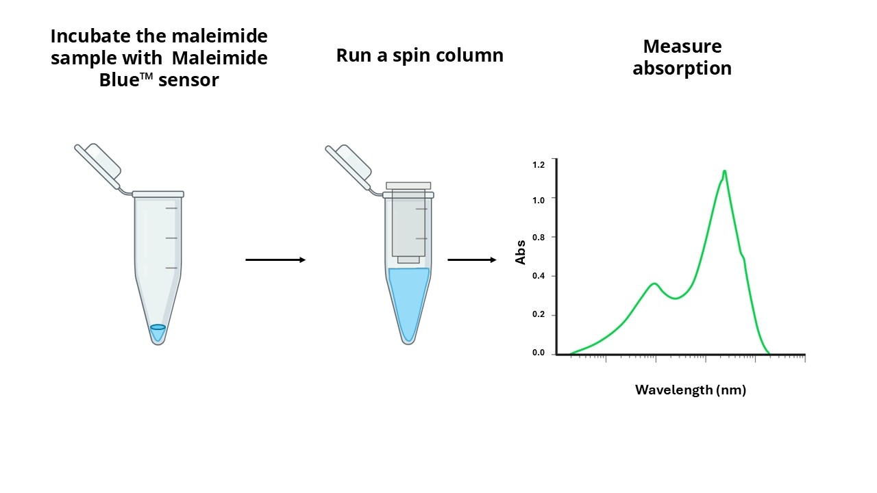
A variety of crosslinking reagents with a maleimide group are widely used for crosslinking proteins to proteins, or proteins to other biomolecules. A key challenge in maleimide crosslinking technology has been the quantitation of maleimide linked to a single protein or antibody. Maleimides can be directly assayed spectrophotometrically at 302 nm. However, the small extinction coefficient of 620 M-1cm-1 renders this assay insensitive, and the assay is further complicated by the protein absorbance at the same wavelength. AAT Bioquest's Amplite® Rapid Colorimetric Maleimide Quantitation Kit provides a rapid and accurate method to quantify maleimide using our proprietary maleimide sensor Maleimide Blue™ with the maximum absorbance at ~780nm. The principle of this assay is that Maleimide Blue™ reacts with the maleimide-linked sample, and the resulted product is run through a single spin column to remove the excess sensor. The absorption spectrum of the purified product is measured, and the amount of maleimide to protein ratio can be calculated from the absorbance ratio of 780 nm and 280 nm (for proteins) or 260 nm (for oligos and nucleic acids). This Amplite® Rapid Maleimide Quantitation kit can be performed in a traditional cuvette, NanoDrop™ Spectrophotometer or a convenient 96-well absorbance plate reader with a UV-transparent plate. This kit has been widely used for the rapid quantification of maleimide group from protein, oligo and nucleic acid samples.

Platform


Absorbance microplate reader

Absorbance900 nm to 250 nm
Recommended plateClear bottom

Components


Example protocol


AT A GLANCE

Important notes
Upon receipt, store Maleimide Blue™ (Component A) at -20°C (prefer at -80°C), kept from light and moisture. When stored properly, the kit components should be stable for six months. Do not freeze Spin Column (Component C). Warm all the components before run the required assays. 50 to 100 µg maleimide-linked antibody or protein sample is needed for determining the amount of maleimide.

SAMPLE EXPERIMENTAL PROTOCOL

Prepare sample solution:

  1. Use 50 to 100 µg maleimide sample (protein or other polymers).

  2. Adjust the volume to 100 µL with Assay Buffer (Component B). Note: The maleimide-linked antibody or protein sample should be in pH = 6.0 buffer and without free maleimide.

Run Maleimide Assay:

  1. Add the maleimide sample to one vial of Maleimide Blue™ (Component A).

  2. Mix them well by repeatedly pipetting for a few times or vortex the vial for a few seconds.

  3. Keep the reaction mixture at room temperature and rotate or shake for 30 - 60 minutes.

Prepare Spin Column for Sample Purification:

  1. Invert the Spin Column (Component C) several times to resuspend the settled gel and remove any bubbles.

  2. Snap off the tip and place the column in the Washing Tube (2 mL, Component D). Remove the cap to allow the excess packing buffer to drain by gravity to the top of the gel bed. If column does not begin to flow, push cap back into column and remove it again to start the flow. Discard the drained buffer, and then place the column back into the Washing Tube. However, centrifuge immediately if the column is placed into a 12 x 75 mm test tube (not provided).

  3. Centrifuge for 1 min in a swinging bucket centrifuge at 1,000 x g (see Centrifugation Notes section) to remove the packing buffer. Discard the buffer.

  4. Apply 1 mL Assay Buffer (Component B) to the column, let the buffer drain out by gravity, or centrifuge the column for 1 min to remove the buffer. Discard the buffer from the collection tube. Repeat this process for 3 - 4 times.

  5. Centrifuge for 2 minutes in a swinging bucket centrifuge at 1,000 x g (see Centrifugation Notes section) to remove the reaction buffer. Discard the buffer. Note: Spin Column (Component C) can fit into 2 mL microcentrifuge tubes or 12 x 75 mm test tubes for sample collection during centrifugation. Use the 2 mL microtubes provided with the columns for the initial column equilibration step. Note: Swinging bucket centrifuges capable of generating a minimum force of 1,000 x g are suitable for Bio-Spin column use. The gravitational force created at a particular revolution speed is a function of the radius of the microcentrifuge rotor. Consult the swinging bucket centrifuge instruction manual for the information about conversion from revolutions per minute (RPM) to centrifugal or g-force. Alternatively, use the equation to calculate the speed in RPM required to reach the gravitational force of 1,000 x g.

    RCF(g) = (1.12 x 10⁻⁵) X (RPM)2 x r 

    RCF = the relative centrifugal force
    RPM = the speed of the rotor
    r = the radius in centimeters measured from the center of the rotor to the middle of the Bio-Spin column

Purify Malemide Reaction Product:

  1. Place the column in a clean Collecting Tube (1.5 mL, Component E). Carefully load the sample (100 µL) directly to the center of the column.

  2. After loading the sample, add 10 µL Assay Buffer (Component B) to the top and centrifuge the column for 5 min at 1,000 x g, and collect the solution into the collecting tube.

Run Absorption Spectra with 0.2mL or 0.5 mL Quartz Cuvette:

  1. Dilute the maleimide reaction product by 5 - 10 folds with Assay Buffer (Component B) depending on the cuvette size used and the absorbance reading. The dilution factor doesn’t affect the final maleimide quantitation result.

  2. Measure the absorption spectrum from 900 nm to 250 nm range, or only read the absorbance number at 280 nm and 782 nm. 

Images


Citations


View all 6 citations: Citation Explorer
Successful acquisition of a neutralizing monoclonal antibody against a novel neutrophil-activating peptide, mitocryptide-1
Authors: Hattori, Tatsuya and Nakashima, Kenta and Marutani, Takayuki and Kiso, Yoshiaki and Nishi, Yoshisuke and Mukai, Hidehito
Journal: Biochemical and biophysical research communications (2015): 54--59
Rapid quantification of maleimide in bioconjuated samples using a novel colorimetric method (TECH2P. 761)
Authors: Liao, Jinfang and Guo, Haitao and Luo, Zhen and Zhao, Qin and Diwu, Jack
Journal: The Journal of Immunology (2015): 206--7
Moderate PEGylation of the carrier protein improves the polysaccharide-specific immunogenicity of meningococcal group A polysaccharide conjugate vaccine
Authors: Zhang, Tingting and Yu, Weili and Wang, Yanfei and Hu, Tao
Journal: Vaccine (2015): 3208--3214
PEG as a spacer arm markedly increases the immunogenicity of meningococcal group Y polysaccharide conjugate vaccine
Authors: Huang, Qingrui and Li, Dongxia and Kang, Aijun and An, Wenqi and Fan, Bei and Ma, Xiaowei and Ma, Guanghui and Su, Zhiguo and Hu, Tao
Journal: Journal of Controlled Release (2013): 382--389
CD133-targeted paclitaxel delivery inhibits local tumor recurrence in a mouse model of breast cancer
Authors: Swaminathan, Suresh Kumar and Roger, Emilie and Toti, Udaya and Niu, Lin and Ohlfest, John R and Panyam, Jayanth
Journal: Journal of Controlled Release (2013): 280--287

References


View all 38 references: Citation Explorer
Characterization of site-specific ScFv PEGylation for tumor-targeting pharmaceuticals
Authors: Natarajan A, Xiong CY, Albrecht H, DeNardo GL, DeNardo SJ.
Journal: Bioconjug Chem (2005): 113
A new maleimide-bound acid-cleavable solid-support reagent for profiling phosphorylation
Authors: Chowdhury SM, Munske GR, Siems WF, Bruce JE.
Journal: Rapid Commun Mass Spectrom (2005): 899
Quantitation of microparticles released from coated-platelets
Authors: Dale GL, Remenyi G, Friese P.
Journal: J Thromb Haemost (2005): 2081
Determination of WR-1065 in human blood by high-performance liquid chromatography following fluorescent derivatization by a maleimide reagent ThioGlo3
Authors: Chen J, Lu Z, Lawrence TS, Smith DE.
Journal: J Chromatogr B Analyt Technol Biomed Life Sci (2005): 161
Reactions of cysteines substituted in the amphipathic N-terminal tail of a bacterial potassium channel with hydrophilic and hydrophobic maleimides
Authors: Li J, Xu Q, Cortes DM, Perozo E, Laskey A, Karlin A.
Journal: Proc Natl Acad Sci U S A (2002): 11605
Resolution, detection, and characterization of redox conformers of human HSF1
Authors: Manalo DJ, Liu AY.
Journal: J Biol Chem (2001): 23554
Site-directed mutation of conserved cysteine residues does not inactivate the Streptococcus pyogenes hyaluronan synthase
Authors: Heldermon CD, Tlapak-Simmons VL, Baggenstoss BA, Weigel PH.
Journal: Glycobiology (2001): 1017
Quantitation of signal transduction
Authors: Krauss S, Br and MD., undefined
Journal: Faseb J (2000): 2581
Determination of S-nitrosoglutathione in human and rat plasma by high-performance liquid chromatography with fluorescence and ultraviolet absorbance detection after precolumn derivatization with o-phthalaldehyde
Authors: Tsikas D, S and mann J, Holzberg D, Pantazis P, Raida M, Frolich JC.
Journal: Anal Biochem (1999): 32
Peroxynitrite modification of protein thiols: oxidation, nitrosylation, and S-glutathiolation of functionally important cysteine residue(s) in the sarcoplasmic reticulum Ca-ATPase
Authors: Viner RI, Williams TD, Schoneich C.
Journal: Biochemistry (1999): 12408