logo
AAT Bioquest

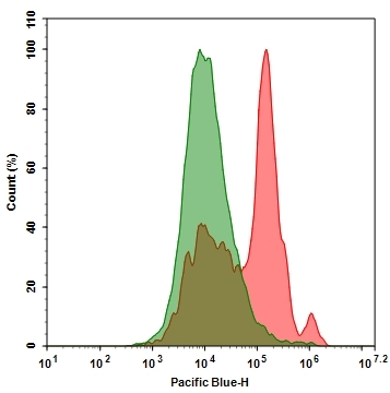
Annexin V, PacBlue conjugate

Annexin V binding assay for identifying apoptosis cells.  Jurkat cells were treated without (Green) or with 1uM staurosporine (Red) at 37 °C for 4-5 hours. Cells were then incubated with Annexin V PacBlue conjugate (Cat#20089) for 30min. The fluorescence signal was monitored using ACEA NovoCyte flow cytometer Pacific Blue channel.
Ordering information
Price
Catalog Number
Unit Size
Quantity
Add to cart
Additional ordering information
Telephone1-800-990-8053
Fax1-800-609-2943
Emailsales@aatbio.com
InternationalSee distributors
Bulk requestInquire
Custom sizeInquire
ShippingStandard overnight for United States, inquire for international
Request quotation
Physical properties
Molecular weight~36000
SolventWater
Spectral properties
Correction Factor (260 nm)0.15
Correction Factor (280 nm)0.2
Extinction coefficient (cm -1 M -1)46000
Excitation (nm)404
Emission (nm)455
Quantum yield0.78
Storage, safety and handling
H-phraseH303, H313, H333
Hazard symbolXN
Intended useResearch Use Only (RUO)
R-phraseR20, R21, R22
StorageFreeze (< -15 °C); Minimize light exposure
UNSPSC12352200

OverviewpdfSDSpdfProtocol


Molecular weight
~36000
Correction Factor (260 nm)
0.15
Correction Factor (280 nm)
0.2
Extinction coefficient (cm -1 M -1)
46000
Excitation (nm)
404
Emission (nm)
455
Quantum yield
0.78
Annexins are a family of proteins that bind to phospholipid membranes in the presence of calcium. Annexin V is a valuable tool for studying cell apoptosis. It is used as a probe to detect cells which have expressed phosphatidylserine on the cell surface, a feature found in apoptosis as well as other forms of cell death. In apoptosis, PS is transferred to the outer leaflet of the plasma membrane. The appearance of phosphatidylserine on the cell surface is a universal indicator of the initial/intermediate stages of cell apoptosis and can be detected before morphological changes can be observed. There are a variety of parameters that can be used for monitoring cell viability. Annexin V-dye conjugates are widely used to monitor cell apoptosis through measuring the translocation of phosphatidylserine (PS). The annexin V, PacBlue conjugate is violet excitable, making it ideal for instruments with a violet laser and for multicolor experiments that include green- or red-fluorescent dyes.

Platform


Flow cytometer

Excitation405 nm laser
Emission450/40 nm filter
Instrument specification(s)Pacific Blue channel

Fluorescence microscope

ExcitationDAPI filter set
EmissionDAPI filter set
Recommended plateBlack wall/clear bottom

Example protocol


AT A GLANCE

Protocol Summary
  1. Prepare cells with test compounds (200 µL/sample).

  2. Add Annexin V conjugate assay solution.

  3. Incubate at room temperature for 30-60 minutes.

  4. Analyze with a flow cytometer or a fluorescence microscope.

Storage and Handling Conditions

The fluorescent annexin V conjugates are stored in a PBS buffer solution containing 0.1% bovine serum albumin (BSA) with a pH of 7.4. To ensure their stability, it is recommended that the solutions be stored at a temperature of -20°C and protected from light. Avoid exposing the fluorescent conjugates to repeated freeze-thaw cycles as this can have a negative effect on their integrity. These solutions can be stored for at least 6 months under the recommended conditions.

SAMPLE EXPERIMENTAL PROTOCOL

Prepare and Incubate Cells with Annexin V Conjugate
  1. Prepare an Annexin V-binding assay buffer: 10 mM HEPES, 140 mM NaCl, and 2.5 mM CaCl2, pH 7.4.

  2. Treat cells with test compounds for a desired period of time (e.g., 4-6 hours for Jurkat cells treated with staurosporine) to induce apoptosis.

  3. Centrifuge the cells to get 1-5×10⁵ cells/tube.

  4. Resuspend cells in 200 μL of the Annexin V-binding assay buffer from Step 1.

  5. Add 2 μL of the Annexin V conjugate to the cells.

    Optional: Add a dead cell stain such as Propidium Iodide (Cat No. 17585) into the cells for necrosis cells.

  6. Incubate at room temperature for 30 to 60 minutes, protected from light.

  7. Add 300 μL of the Annexin V-binding assay buffer (from Step 1) to increase volume before analyzing the cells with
    a flow cytometer or fluorescence microscope.

  8. Monitor the fluorescence intensity by using a flow cytometer or a fluorescence microscope.

Flow Cytometer Protocol
  1. Quantify Annexin V conjugates binding by using a flow cytometer with appropriate filters.

    Note: It is not common to perform Annexin V binding flow cytometric analysis on adherent cells due to the possibility of membrane damage during cell detachment or harvesting. However, previous studies by Casiola-Rosen et al. and van Engelend et al. (refer to Refs 1 and 2) have demonstrated methods for using Annexin V in flow cytometry on adherent cell types.

Fluorescence Microscope Protocol
  1. Pipette the cell suspension from Step 6, rinse 1-2 times with Annexin V-binding assay buffer (Step 1), and then resuspend the cells with the Annexin V-binding assay buffer (Step 1).

  2. Add the cells on a glass slide that is covered with a glass cover slip.

    Note: For adherent cells, it is recommended to grow the cells directly on a cover slip. 

  3. After incubation with Annexin V conjugate (Step 6), rinse 1-2 times with Annexin V-binding assay buffer (Step 1), and add
    Annexin V-binding assay buffer (Step 1) back to the cover slip.

  4. Invert the cover slip on a glass slide and visualize the cells. The cells can also be fixed in 2% formaldehyde after incubation with Annexin V conjugate and visualized under a microscope with the appropriate filter set.

APPENDIX

References
  1. Pascal Clerc, Pauline Jeanjean, Nicolas Halalli, Michel Gougeon, Bernard Pipy, Julian Carrey, Daniel Fourmy, Véronique Gigoux. Journal of Controlled Release (2017).

  2. Hanshaw RG, Lakshmi C, Lambert TN, Johnson JR, Smith BD. Chembiochem, 6, 2214. (2005).

Spectrum


Open in Advanced Spectrum Viewer
spectrum

Spectral properties

Correction Factor (260 nm)0.15
Correction Factor (280 nm)0.2
Extinction coefficient (cm -1 M -1)46000
Excitation (nm)404
Emission (nm)455
Quantum yield0.78

Product Family


NameExcitation (nm)Emission (nm)Extinction coefficient (cm -1 M -1)Quantum yieldCorrection Factor (260 nm)Correction Factor (280 nm)
Annexin V-Cy3 conjugate55556915000010.1510.070.073
Annexin V-Cy5 conjugate65667025000010.2510.030.03
Annexin V-Cy7 conjugate7567792500000.30.050.036
Annexin V, XFD350 conjugate *XFD350 Same Structure to Alexa Fluor™ 350*34344119000-0.250.19
Annexin V, XFD488 conjugate *XFD488 Same Structure to Alexa Fluor™ 488*499520710000.9210.300.11
Annexin V, XFD594 conjugate *XFD594 Same Structure to Alexa Fluor™ 594*590618920000.6610.430.56

Images


Citations


View all 2 citations: Citation Explorer
Targeted Magnetic Intra-Lysosomal Hyperthermia produces lysosomal reactive oxygen species and causes Caspase-1 dependent cell death
Authors: Clerc, Pascal and Jeanjean, Pauline and Halalli, Nicolas and Gougeon, Michel and Pipy, Bernard and Carrey, Julian and Fourmy, Daniel and Gigoux, V&eacute;ronique
Journal: Journal of Controlled Release (2017)

References


View all 32 references: Citation Explorer
Gold fluorescent annexin A5 as a novel apoptosis detection tool
Authors: Kurschus FC, Pal PP, Baumler P, Jenne DE, Wiltschi B, Budisa N.
Journal: Cytometry A (2009): 626
Glycogen synthase kinase-3 and Omi/HtrA2 induce annexin A2 cleavage followed by cell cycle inhibition and apoptosis
Authors: Wang CY, Lin YS, Su WC, Chen CL, Lin CF.
Journal: Mol Biol Cell (2009): 4153
Evaluation of annexin V and Calcein-AM as markers of mononuclear cell apoptosis during human immunodeficiency virus infection
Authors: Palma PF, Baggio GL, Spada C, Silva RD, Ferreira SI, Treitinger A.
Journal: Braz J Infect Dis (2008): 108
Detection of apoptosis induced by new type gosling viral enteritis virus in vitro through fluorescein annexin V-FITC/PI double labeling
Authors: Chen S, Cheng AC, Wang MS, Peng X.
Journal: World J Gastroenterol (2008): 2174
Measurement of annexin V uptake and lactadherin labeling for the quantification of apoptosis in adherent Tca8113 and ACC-2 cells
Authors: Hu T, Shi J, Jiao X, Zhou J, Yin X.
Journal: Braz J Med Biol Res (2008): 750
Two-way trafficking of Annexin V positive cells between mother and fetus: determination of apoptosis at delivery
Authors: Kolialexi A, Tsangaris GT, Anagnostopoulos A, Chondros D, Bagiokos V, Kitsiou S, Kanavakis E, Mavrou A.
Journal: Prenat Diagn (2007): 348
Rhodamine B isothiocyanate doped silica-coated fluorescent nanoparticles (RBITC-DSFNPs)-based bioprobes conjugated to Annexin V for apoptosis detection and imaging
Authors: Shi H, He X, Wang K, Yuan Y, Deng K, Chen J, Tan W.
Journal: Nanomedicine (2007): 266
Annexin A5-functionalized bimodal lipid-based contrast agents for the detection of apoptosis
Authors: van Tilborg GA, Mulder WJ, Deckers N, Storm G, Reutelingsperger CP, Strijkers GJ, Nicolay K.
Journal: Bioconjug Chem (2006): 741
Analysis of cycloheximide-induced apoptosis in human leukocytes: Fluorescence microscopy using annexin V/propidium iodide versus acridin orange/ethidium bromide
Authors: Baskic D, Popovic S, Ristic P, Arsenijevic NN.
Journal: Cell Biol Int (2006): 924
Quantum dots based probes conjugated to annexin V for photostable apoptosis detection and imaging
Authors: Le Gac S, Vermes I, van den Berg A.
Journal: Nano Lett (2006): 1863