logo
AAT Bioquest

Annexin V

FITC Annexin V is a highly fluorescent conjugate used to identify and quantitate apoptotic cells by flow cytometry.

Product Description

Annexin V is a 35-36 kDa phospholipid-binding protein that has a high affinity for phosphatidylserine (PS) residues. In apoptotic cells, PS is translocated from the inner to the outer leaflet of the plasma membrane, making it accessible to Annexin V conjugated to fluorescein isothiocyanate (FITC). By binding to PS, Annexin V FITC can be used to detect and quantify apoptotic cells via flow cytometry or fluorescence microscopy.

Key Features

  • High Sensitivity and Specificity to efficiently detect early apoptotic cells by binding to exposed PS.
  • Dual Staining Capability can be used in conjunction with propidium iodide (PI) to distinguish between apoptotic and necrotic cells.
  • Non-Radioactive Assay provides a safe and easy method for apoptosis detection without the need for radioactive materials.
  • Rapid and Simple Protocol enables minimal hands-on time with straightforward staining and analysis procedures.
  • Versatile Applications suitable for use in various cell types and experimental conditions.

Mechanism of Action

During apoptosis, the integrity of the plasma membrane changes, leading to the translocation of phosphatidylserine (PS) from the inner to the outer leaflet. Annexin V FITC, a fluorophore-conjugated protein with a high affinity for PS, selectively binds to the externalized PS residues. This binding event is detectable and quantifiable via flow cytometry or fluorescence microscopy. The intensity of the FITC signal enables precise identification and quantification of apoptotic cells, as it directly correlates with Annexin V binding to PS on the cell surface.
Example protocol

AT A GLANCE

Protocol Summary
  1. Prepare cells with test compounds (200 µL/sample).

  2. Add Annexin V conjugate assay solution.

  3. Incubate at room temperature for 30-60 minutes.

  4. Analyze with a flow cytometer or a fluorescence microscope.

Storage and Handling Conditions

The fluorescent annexin V conjugates are stored in a PBS buffer solution containing 0.1% bovine serum albumin (BSA) with a pH of 7.4. To ensure their stability, it is recommended that the solutions be stored at a temperature of -20°C and protected from light. Avoid exposing the fluorescent conjugates to repeated freeze-thaw cycles as this can have a negative effect on their integrity. These solutions can be stored for at least 6 months under the recommended conditions.

SAMPLE EXPERIMENTAL PROTOCOL

Prepare and Incubate Cells with Annexin V Conjugate
  1. Prepare an Annexin V-binding assay buffer: 10 mM HEPES, 140 mM NaCl, and 2.5 mM CaCl2, pH 7.4.

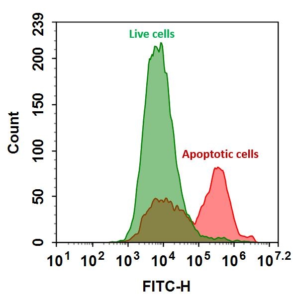
  2. Treat cells with test compounds for a desired period of time (e.g., 4-6 hours for Jurkat cells treated with staurosporine) to induce apoptosis.

  3. Centrifuge the cells to get 1-5×10⁵ cells/tube.

  4. Resuspend cells in 200 μL of the Annexin V-binding assay buffer from Step 1.

  5. Add 2 μL of the Annexin V conjugate to the cells.

    Optional: Add a dead cell stain such as Propidium Iodide (Cat No. 17585) into the cells for necrosis cells.

  6. Incubate at room temperature for 30 to 60 minutes, protected from light.

  7. Add 300 μL of the Annexin V-binding assay buffer (from Step 1) to increase volume before analyzing the cells with
    a flow cytometer or fluorescence microscope.

  8. Monitor the fluorescence intensity by using a flow cytometer or a fluorescence microscope.

Flow Cytometer Protocol
  1. Quantify Annexin V conjugates binding by using a flow cytometer with appropriate filters.

    Note: It is not common to perform Annexin V binding flow cytometric analysis on adherent cells due to the possibility of membrane damage during cell detachment or harvesting. However, previous studies by Casiola-Rosen et al. and van Engelend et al. (refer to Refs 1 and 2) have demonstrated methods for using Annexin V in flow cytometry on adherent cell types.

Fluorescence Microscope Protocol
  1. Pipette the cell suspension from Step 6, rinse 1-2 times with Annexin V-binding assay buffer (Step 1), and then resuspend the cells with the Annexin V-binding assay buffer (Step 1).

  2. Add the cells on a glass slide that is covered with a glass cover slip.

    Note: For adherent cells, it is recommended to grow the cells directly on a cover slip. 

  3. After incubation with Annexin V conjugate (Step 6), rinse 1-2 times with Annexin V-binding assay buffer (Step 1), and add
    Annexin V-binding assay buffer (Step 1) back to the cover slip.

  4. Invert the cover slip on a glass slide and visualize the cells. The cells can also be fixed in 2% formaldehyde after incubation with Annexin V conjugate and visualized under a microscope with the appropriate filter set.

APPENDIX

References
  1. Pascal Clerc, Pauline Jeanjean, Nicolas Halalli, Michel Gougeon, Bernard Pipy, Julian Carrey, Daniel Fourmy, Véronique Gigoux. Journal of Controlled Release (2017).

  2. Hanshaw RG, Lakshmi C, Lambert TN, Johnson JR, Smith BD. Chembiochem, 6, 2214. (2005).

Spectrum

Product family

NameExcitation (nm)Emission (nm)Extinction coefficient (cm -1 M -1)Correction Factor (260 nm)Correction Factor (280 nm)
Annexin V, TRITC Labeled5445701000000.270.34

Citations

View all 3 citations: Citation Explorer
Galectin-1 overexpression induces normal fibroblasts translate into cancer-associated fibroblasts and attenuates the sensitivity of anlotinib in lung cancer
Authors: Zhang, Lei and Chen, Wenbang and Li, Xiaojun and Wang, Gengming and Xing, Fubao and Zhu, Xiao
Journal: Cell Adhesion \& Migration (2024): 1--11
The role of non-coding RNA ASBEL regulation of BTG3 in pancreatic ductal adenocarcinoma tumor progression
Authors: Chen, Jing and Zhu, Ming-Yuan and Huang, Yan-Hua and Ling, Yi-Ting and Gu, Tian-Yuan and Zhou, Quan and Fei, Ming-Jian and Zhou, Zhong-Cheng
Journal: Current Therapeutic Research (2023): 100700
Response of Mammalian cells to non-thermal intense narrowband pulsed electric fields
Authors: Katsuki, Sunao and Li, Yulan and Miyakawa, Daiki and Yamada, Ryo and Onishi, Nobuaki and Lim, Soowon
Journal: (2017): 1358--1361

References

View all 90 references: Citation Explorer
Non-fusion expression in Escherichia coli: Single-step purification of recombinant human annexin A5 for detection of apoptosis
Authors: Wang F, He XW, Yan HL, Huang JJ, Zhang Y, Jiang L, Gao YJ, Sun SH.
Journal: Protein Expr Purif (2006): 80
PET imaging of apoptosis with (64)Cu-labeled streptavidin following pretargeting of phosphatidylserine with biotinylated annexin-V
Authors: Cauchon N, Langlois R, Rousseau JA, Tessier G, Cadorette J, Lecomte R, Hunting DJ, Pavan RA, Zeisler SK, van Lier JE.
Journal: Eur J Nucl Med Mol Imaging. (2006)
The impact of altered annexin I protein levels on apoptosis and signal transduction pathways in prostate cancer cells
Authors: Hsiang CH, Tunoda T, Whang YE, Tyson DR, Ornstein DK.
Journal: Prostate (2006): 1413
Lymphoma cells protected from apoptosis by dysregulated bcl-2 continue to bind annexin V in response to B-cell receptor engagement: a cautionary tale
Authors: Holder MJ, Barnes NM, Gregory CD, Gordon J.
Journal: Leuk Res (2006): 77
Quantum dots based probes conjugated to annexin V for photostable apoptosis detection and imaging
Authors: Le Gac S, Vermes I, van den Berg A.
Journal: Nano Lett (2006): 1863
Page updated on October 8, 2024

Ordering information

Price
Conjugate
Unit size
100 tests
300 tests
Catalog Number
Quantity
Add to cart

Additional ordering information

Telephone1-800-990-8053
Fax1-800-609-2943
Emailsales@aatbio.com
InternationalSee distributors
Bulk requestInquire
Custom sizeInquire
Technical SupportContact us
Purchase orderSend to sales@aatbio.com
ShippingStandard overnight for United States, inquire for international
Request quotation

Physical properties

Molecular weight

~36000

Solvent

Water

Spectral properties

Correction Factor (280 nm)

0.35

Extinction coefficient (cm -1 M -1)

73000

Excitation (nm)

491

Emission (nm)

516

Quantum yield

0.92

Storage, safety and handling

H-phraseH303, H313, H333
Hazard symbolXN
Intended useResearch Use Only (RUO)
R-phraseR20, R21, R22

Storage

Freeze (< -15 °C); Minimize light exposure
UNSPSC12352200

Platform

Flow cytometer

Excitation488 nm laser
Emission530, 30 nm filter
Instrument specification(s)FITC channel

Fluorescence microscope

ExcitationFITC filter set
EmissionFITC filter set
Recommended plateBlack wall, clear bottom
Binding activity of FITC-Annexin V conjugate to phosphatidylserine (PS) residues in Jurkat cells. Jurkat cells were treated without (Green) or with 1 μM staurosporine (Red) at 37°C for 4 hours and then labeled with FITC-Annexin V conjugate for 30 minutes. The fluorescence intensity was measured using an ACEA NovoCyte flow cytometer in the FITC channel.