Products
Services
Resources
Selection Guides
About
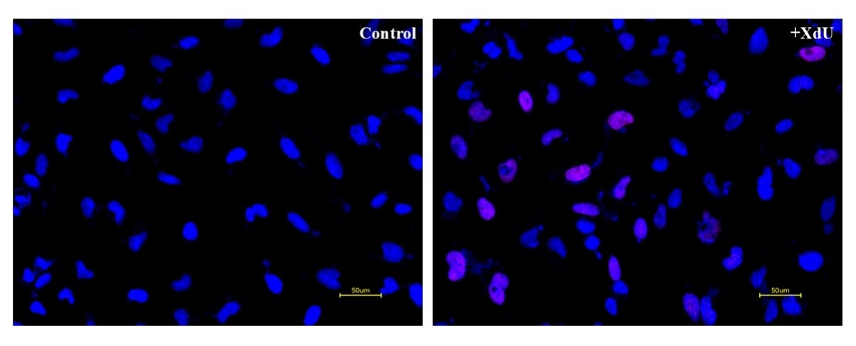
Bucculite™ XdU Cell Proliferation Fluorescence Imaging Kit
Red Fluorescence
Monitoring cell proliferation is one of the most reliable methods to assess cell viability, cell cycles and genotoxicity. An essential way to detect cell proliferation is to measure DNA synthesis in the presence of thymidine during the S-phase of cells growth. Bucculite™ XdU Cell Proliferation Fluorescence Imaging Kit uses XdU which is incorporated into cellular DNA during DNA synthesis. After fixing cells, the incorporated XdU is labelled with iFluor® 555 MTA. The resulted iFluor® 555-labeled DNA formed in cells is imaged with a Cy3 filter set. Bucculite™ XdU Cell Proliferation Fluorescence Imaging Kit provides an alternative to anti-BrdU antibody-based assay and EdU click chemistry-based assay. It is sensitive and might be used for measuring active DNA synthesis at single-cell level.
| Catalog | Size | Price | Quantity |
|---|---|---|---|
| 22327 | 200 Tests | Price |
Usage and storage
| Intended use | Research Use Only (RUO) |
Instrument settings
| Fluorescence microscope | |
| Excitation | Cy3/TRITC filter set |
| Emission | Cy3/TRITC filter set |
| Recommended plate | Black wall/clear bottom |
Contact us
| Telephone | |
| Fax | |
| sales@aatbio.com | |
| International | See distributors |
| Bulk request | Inquire |
| Custom size | Inquire |
| Technical Support | Contact us |
| Request quotation | Request |
| Purchase order | Send to sales@aatbio.com |
| Shipping | Standard overnight for United States, inquire for international |
Page updated on July 26, 2026