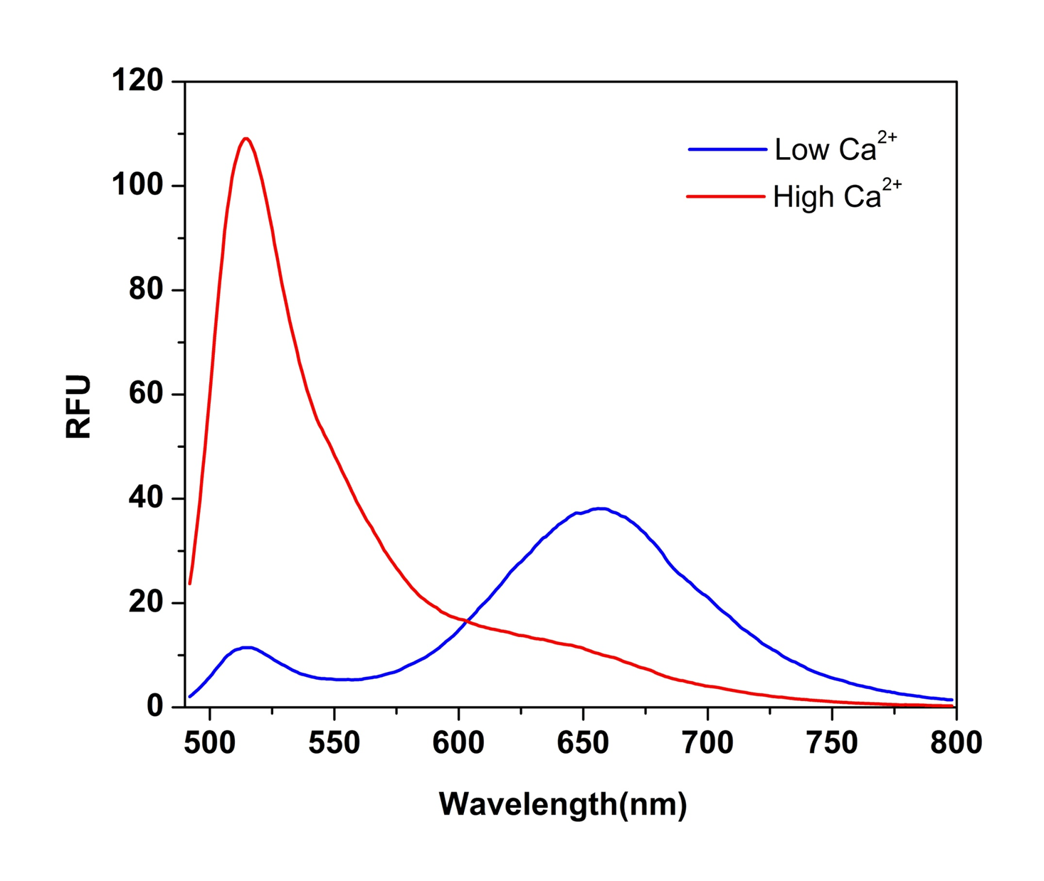
Cal Red™ R525/650 potassium salt
Price | |
Catalog Number | |
Unit Size | |
Quantity |
Telephone | 1-800-990-8053 |
Fax | 1-800-609-2943 |
sales@aatbio.com | |
International | See distributors |
Bulk request | Inquire |
Custom size | Inquire |
Shipping | Standard overnight for United States, inquire for international |
Dissociation constant (Kd, nM) | 330 |
Molecular weight | ~1000 |
Solvent | Water |
H-phrase | H303, H313, H333 |
Hazard symbol | XN |
Intended use | Research Use Only (RUO) |
R-phrase | R20, R21, R22 |
Storage | Freeze (< -15 °C); Minimize light exposure |
UNSPSC | 12352200 |
Overview | ![]() ![]() |
Molecular weight ~1000 | Dissociation constant (Kd, nM) 330 |
Example protocol
SAMPLE EXPERIMENTAL PROTOCOL
Calcium calibration can be carried out by measuring the fluorescence intensity of the salt form (25 to 50 µM in
fluorescence microplate readers) of the indicators in solutions with precisely known free Ca2+ concentrations. Calibration
solutions can be used based on 30 mM MOPS EGTA Ca2+ buffer. In general, water contains trace amount of calcium ion. It
is highly recommended to use 30 mM MOPS + 100 mM KCl, pH 7.2 as buffer system. One can simply make a 0 and 39 µM
calcium stock solutions as listed below, and these 2 solutions are used to make a serial solution of different Ca2+
concentrations.
A. 0 µM calcium: 30 mM MOPS + 100 mM KCl, pH 7.2 buffer + 10 mM EGTA
B. 39 µM calcium: 30 mM MOPS + 100 mM KCl, pH 7.2 buffer + 10 mM EGTA + 10 mM CaCl2
To determine either the free calcium concentration of a solution or the Kd of a single-wavelength calcium indicator,
the following equation is used:
[Ca]free = Kd[F ─ Fmin]/[Fmax ─ F]
Where F is the fluorescence intensity of the indicator at a specific experimental calcium level, Fmin is the
fluorescence intensity in the absence of calcium and Fmax is the fluorescence intensity of the calciumsaturated probe.
The dissociation constant (Kd) is a measure of the affinity of the probe for calcium. The calcium-binding and
spectroscopic properties of fluorescent indicators vary quite significantly in cellular environments compared to calibration
solutions. In situ response calibrations of intracellular indicators typically yield Kd values significantly higher than in vitro
determinations. In situ calibrations are performed by exposing loaded cells to controlled Ca2+ buffers in the presence of
ionophores such as A-23187, 4-bromo A-23187 and ionomycin. Alternatively, cell permeabilization agents such as digitonin
or Triton® X-100 can be used to expose the indicator to the controlled Ca2+ levels of the extracellular medium.
Images
References
Authors: Wendt ER, Ferry H, Greaves DR, Keshav S.
Journal: PLoS One (2015): e0119532
Authors: Bailey S, Macardle PJ.
Journal: J Immunol Methods (2006): 220
Authors: Walczysko P, Wagner E, Albrechtova JT.
Journal: Cell Calcium (2000): 23
Authors: Su ZL, Li N, Sun YR, Yang J, Wang IM, Jiang SC.
Journal: Shi Yan Sheng Wu Xue Bao (1998): 323
Authors: Blackwood AM, Sagnella GA, Mark and u ND, MacGregor GA.
Journal: J Hum Hypertens (1997): 601
Authors: Wu Y, Clusin WT.
Journal: Am J Physiol (1997): H2161
Authors: Floto RA, Mahaut-Smith MP, Somasundaram B, Allen JM.
Journal: Cell Calcium (1995): 377
Authors: Schild D, Jung A, Schultens HA.
Journal: Cell Calcium (1994): 341
Authors: Novak EJ, Rabinovitch PS.
Journal: Cytometry (1994): 135
Authors: Lew VL, Etzion Z, Bookchin RM, daCosta R, Vaananen H, Sassaroli M, Eisinger J.
Journal: Biochim Biophys Acta (1993): 152
Application notes
A Meta-Analysis of Common Calcium Indicators
A New Red Fluorescent & Robust Screen Quest™ Rhod-4™ Ca2+Indicator for Screening GPCR & Ca2+ Channel Targets
A New Robust No-Wash FLIPR Calcium Assay Kit for Screening GPCR and Calcium Channel Targets
A Novel NO Wash Probeniceid-Free Calcium Assay for Functional Analysis of GPCR and Calcium Channel Targets
FAQ
Are there upgraded trypan blue derivatives for cell viability testing?
Can I intracellularly measure mitochondria calcium flux and changes in mitochondria membrane potential at the same time?
Do you offer any products for measuring intracellular calcium concentration or movement by flow cytometry?
Does EDTA inactivate proteinase K?