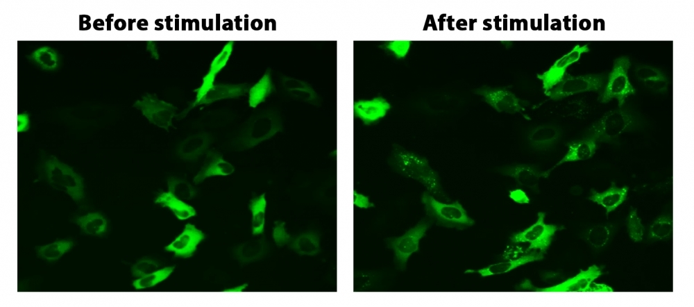
Cell Meter™ Beta-Arrestin Translocation GPCR Signaling Kit
Virtually all G protein coupled receptors (GPCRs) rapidly undergo desensitization by a common pathway upon activation by ligand binding. The binding of beta-arrestin, a cytoplasmic protein, to an activated receptor deactivates the GPCR signaling and initiates the translocation of the receptor into the cell where the ligand is removed, and the receptor is recycled back to the cell membrane. By attaching a fluorescent label, such as GFP, to beta-arrestin, the location of the receptor arrestin complex can be monitored. Since desensitization only occurs with an activated receptor, monitoring beta-arrestin translocation and subsequent receptor recycling provides a reliable method to detect the activation of a GPCR target. Cell Meter™ Beta-Arrestin translocation GPCR Signaling Kit provides a powerful functional assay to screen activities of target compounds against known or orphan GPCR targets via fluorescence imaging. The activation of the targeted GPCR induces the translocation of the fluorescence to the cell membrane and/or to endocytic vesicles.
![Translocation of beta-arrestin in HeLa cells. HeLa cells were transiently transfected with beta-arrestin-GFP and vasopressin receptor 2 (V2R). HeLa cells were cultured in a 6-well plate and grown to ~60% confluence. Equal amounts of beta-arrestin-GFP (1.5 µg) and V2R plasmids (1.5 µg) were transfected with 9 µL of Transfectamine™ 5000. Cells were transferred to a 96-well plate ~ 30 hours after transfection. Vasopressin (1 µM) was added to the cells ~ 48 hours after transfection to induce beta-arrestin-GFP translocation. Images were taken before and 2 hours after the vasopressin treatment under a fluorescent microscope using the FITC channel.](/_next/image?url=https%3A%2F%2Fimages.aatbio.com%2Fproducts%2Ffigures-and-data%2Fcell-meter-beta-arrestin-translocation-gpcr-signaling-kit%2Ffigure-for-cell-meter-beta-arrestin-translocation-gpcr-signaling-kit_6tnFs.jpg&w=640&q=75)
![Translocation of beta-arrestin in HeLa cells. HeLa cells were transiently transfected with beta-arrestin-GFP and vasopressin receptor 2 (V2R). HeLa cells were cultured in a 6-well plate and grown to ~60% confluence. Equal amounts of beta-arrestin-GFP (1.5 µg) and V2R plasmids (1.5 µg) were transfected with 9 µL of Transfectamine™ 5000. Cells were transferred to a 96-well plate ~ 30 hours after transfection. Vasopressin (1 µM) was added to the cells ~ 48 hours after transfection to induce beta-arrestin-GFP translocation. Images were taken before and 2 hours after the vasopressin treatment under a fluorescent microscope using the FITC channel.](/_next/image?url=https%3A%2F%2Fimages.aatbio.com%2Fproducts%2Ffigures-and-data%2Fcell-meter-beta-arrestin-translocation-gpcr-signaling-kit%2Ffigure-for-cell-meter-beta-arrestin-translocation-gpcr-signaling-kit_6tnFs.jpg&w=128&q=25)
Example protocol
AT A GLANCE
Protocol summary
- Prepare cells for transfection
- Prepare Transfectamine™ 5000-DNA mixture
- Add Transfectamine™ 5000-DNA mixture to cell culture, and incubate overnight
- Transfer the transfected cells to a 96-well plate 24-30 hours after transfection, and incubate the culture overnight
- Analyze translocation induced by GPCR activation under a fluorescence microscope
CELL PREPARATION
- Seed the cells at a density such that they will be ~60-70% confluent at the time of transfection.
- Replace with fresh growth medium before transfection.
Note For example, replace with 2 mL of medium per well for 6-well plates and 6 mL of medium for 10 cm plates.
PREPARATION OF STOCK SOLUTIONS
Unless otherwise noted, all unused stock solutions should be divided into single-use aliquots and stored at -20 °C after preparation. Avoid repeated freeze-thaw cycles.
beta-arrestin-GFP DNA stock solution
Add 10 µL of ddH2O to the vial of beta-arrestin-GFP DNA (Component A), mix well to have the final concentration of 1 µg/µL.PREPARATION OF WORKING SOLUTION
- Mix 3 µg of DNA [for example, 1.5 µg of Beta-arrestin-GFP DNA stock solution and 1.5 µg DNA of the GPCR that you are interested] with 200 µL of serum-free medium.
- Add 9 µL of Transfectamine™ 5000 (Component B) to the mixture.
- Mix well and incubate at room temperature for 20 minutes.
Note The ratio of Transfectamine™ 5000 and DNA need to be optimized for different cell lines, in general, in our testings, the ratio for Transfectamine™ 5000 Transfection Reagent (µL) to DNA (µg) Ratio should be 3-5 µL : 1 µg.
Component | 6-well plate (per well) | 10 cm plate |
Fresh culture medium | 2 mL | 6 mL |
Plasmid | 3 µg | 10 µg |
Serum-free medium | 200 µL | 600 µL |
Transfectamine™ 5000 Transfection Reagent | ~9 µL | ~30 µL |
SAMPLE EXPERIMENTAL PROTOCOL
Transfection and Translocation protocol
- Add Transfectamine™ 5000 -DNA mixture to the culture plate and incubate overnight.
Note The recombinant protein can start to be detected as early as 16 hours after transfection. The maximal expression level may be observed 72~96 hours after transfection. - Transfer the transfected cells to a 96-well plate 24-30 hours after transfection and incubate overnight.
- Monitor the beta-arrestin translocation induced by the receptor activation under a fluorescence microscope with the FITC filter (Ex/Em = 488/530 nm).
References
View all 10 references: Citation Explorer
Screening cellular feature measurements for image-based assay development.
Authors: Logan, David J and Carpenter, Anne E
Journal: Journal of biomolecular screening (2010): 840-6
Authors: Logan, David J and Carpenter, Anne E
Journal: Journal of biomolecular screening (2010): 840-6
Kappa opioid receptor screen with the Tango beta-arrestin recruitment technology and characterization of hits with second-messenger assays.
Authors: Doucette, Christopher and Vedvik, Kevin and Koepnick, Elizabeth and Bergsma, Aaron and Thomson, Brian and Turek-Etienne, Tammy C
Journal: Journal of biomolecular screening (2009): 381-94
Authors: Doucette, Christopher and Vedvik, Kevin and Koepnick, Elizabeth and Bergsma, Aaron and Thomson, Brian and Turek-Etienne, Tammy C
Journal: Journal of biomolecular screening (2009): 381-94
Multiplexed assays by high-content imaging for assessment of GPCR activity.
Authors: Ross, D A and Lee, S and Reiser, V and Xue, J and Alves, K and Vaidya, S and Kreamer, A and Mull, R and Hudak, E and Hare, T and Detmers, P A and Lingham, R and Ferrer, M and Strulovici, B and Santini, F
Journal: Journal of biomolecular screening (2008): 449-55
Authors: Ross, D A and Lee, S and Reiser, V and Xue, J and Alves, K and Vaidya, S and Kreamer, A and Mull, R and Hudak, E and Hare, T and Detmers, P A and Lingham, R and Ferrer, M and Strulovici, B and Santini, F
Journal: Journal of biomolecular screening (2008): 449-55
The ligand-independent translocation assay: an enabling technology for screening orphan G protein-coupled receptors by arrestin recruitment.
Authors: Oakley, Robert H and Hudson, Christine C and Sjaastad, Michael D and Loomis, Carson R
Journal: Methods in enzymology (2006): 50-63
Authors: Oakley, Robert H and Hudson, Christine C and Sjaastad, Michael D and Loomis, Carson R
Journal: Methods in enzymology (2006): 50-63
High-content screening of known G protein-coupled receptors by arrestin translocation.
Authors: Hudson, Christine C and Oakley, Robert H and Sjaastad, Michael D and Loomis, Carson R
Journal: Methods in enzymology (2006): 63-78
Authors: Hudson, Christine C and Oakley, Robert H and Sjaastad, Michael D and Loomis, Carson R
Journal: Methods in enzymology (2006): 63-78