Products
Services
Resources
Selection Guides
About
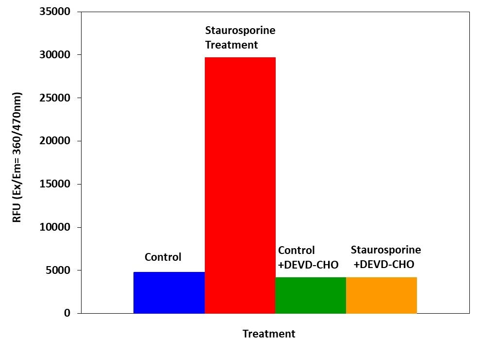
Cell Meter™ Caspase 3/7 Activity Apoptosis Assay Kit
Blue Fluorescence
Our Cell Meter™ assay kits are a set of tools for monitoring cell viability. There are a variety of parameters that can be used for monitoring cell viability. This particular kit is designed to monitor cell apoptosis through measuring Caspase 3 activation. Caspase 3 is widely accepted as a reliable indicator for cell apoptosis since the activation of caspase-3 (CPP32/apopain) is important for the initiation of apoptosis. Caspase 3 has substrate selectivity for the peptide sequence Asp-Glu-Val-Asp (DEVD). This kit uses Ac-DEVD-AMC as a fluorogenic indicator for caspase-3 activity. Cleavage of AMC peptides by caspase 3 generates strongly fluorescent AMC that is monitored fluorimetrically at 450-480 nm with excitation of 340-370 nm. The kit provides all the essential components with an optimized assay protocol. The assay is robust, and can be readily adapted for high-throughput assays. Using 100 uL of reagents per well in a 96-well format, this kit provides sufficient reagents to perform 200 assays. Using 25 uL of reagents per well in a 384-well format, this kit provides sufficient reagents to perform 800 assays.
| Catalog | Size | Price | Quantity |
|---|---|---|---|
| 22795 | 200 Tests | Price |

Spectral properties
| Excitation (nm) | 341 |
| Emission (nm) | 441 |
Usage and storage
| Intended use | Research Use Only (RUO) |
Instrument settings
| Fluorescence microplate reader | |
| Excitation | 360 nm |
| Emission | 470 nm |
| Cutoff | 420 nm |
| Recommended plate | Black wall/clear bottom |
| Instrument specification(s) | Top/Bottom read mode |
Contact us
| Telephone | |
| Fax | |
| sales@aatbio.com | |
| International | See distributors |
| Bulk request | Inquire |
| Custom size | Inquire |
| Technical Support | Contact us |
| Request quotation | Request |
| Purchase order | Send to sales@aatbio.com |
| Shipping | Standard overnight for United States, inquire for international |
Page updated on July 16, 2026