logo
AAT Bioquest

Cell Meter™ Caspase 3/7 Activity Apoptosis Assay Kit *Blue Fluorescence*

Our Cell Meter™ assay kits are a set of tools for monitoring cell viability. There are a variety of parameters that can be used for monitoring cell viability. This particular kit is designed to monitor cell apoptosis through measuring Caspase 3 activation. Caspase 3 is widely accepted as a reliable indicator for cell apoptosis since the activation of caspase-3 (CPP32/apopain) is important for the initiation of apoptosis. Caspase 3 has substrate selectivity for the peptide sequence Asp-Glu-Val-Asp (DEVD). This kit uses Ac-DEVD-AMC as a fluorogenic indicator for caspase-3 activity. Cleavage of AMC peptides by caspase 3 generates strongly fluorescent AMC that is monitored fluorimetrically at 450-480 nm with excitation of 340-370 nm. The kit provides all the essential components with an optimized assay protocol. The assay is robust, and can be readily adapted for high-throughput assays. Using 100 uL of reagents per well in a 96-well format, this kit provides sufficient reagents to perform 200 assays. Using 25 uL of reagents per well in a 384-well format, this kit provides sufficient reagents to perform 800 assays.
Example protocol

AT A GLANCE

Protocol Summary
  1. Prepare cells with test compounds (100 µL/well/96-well plate or 25 µL/well/384-well plate).

  2. Add an equal volume of Caspase 3/7 Substrate working solution.

  3. Incubate at room temperature for 1 hour.

  4. Monitor fluorescence intensity at Ex/Em = 360/470 nm (Cutoff = 420 nm).

Important Note

Thaw one vial of each kit component at room temperature before starting the experiment.

CELL PREPARATION

For guidelines on cell sample preparation, please visit:

https://www.aatbio.com/resources/guides/cell-sample-preparation.html

PREPARATION OF WORKING SOLUTION

  1. Prepare a Caspase 3/7 Substrate working solution by combining 50 µL of Caspase 3/7 Substrate (Component A) with 10 mL of Assay Buffer (Component B), and mix well.

    Note: Aliquot and store any unused Components A and B at -20°C. Avoid repeated freeze/thaw cycles.

SAMPLE EXPERIMENTAL PROTOCOL

  1. To treat cells, add 10 µL per well of 10X test compounds for a 96-well plate or 5 µL per well of 5X test compounds for a 384-well plate into PBS or the desired buffer. For blank wells containing only medium, add an equivalent volume of the compound buffer.

  2. Incubate the cell plate at 37°C with 5% CO2 for a desired duration to induce apoptosis (e.g., 4 to 6 hours for Jurkat cells treated with camptothecin). Adjust the time as necessary based on the cell type and treatment conditions.

  3. Add 100 µL/well for a 96-well plate or 25 µL/well for a 384-well plate of Caspase 3/7 Substrate working solution.

  4. Incubate the plate at room temperature for at least 1 hour, protected from light.

    Note: To confirm the inhibition of the caspase 3/7-like activities, add 1 µL of 1 mM Ac-DEVD-CHO caspase 3/7 inhibitor to selected samples 10 minutes before adding the Caspase 3/7 working solution at room temperature.

  5. Centrifuge cell plate (especially for the non-adherent cells) at 800 rpm for 2 minutes (brake off).

  6. Monitor the fluorescence intensity with a fluorescence microplate reader at Ex/Em = 360/470 nm (Cutoff = 420 nm).

Spectrum

Product family

Citations

View all 38 citations: Citation Explorer
High-dose ascorbate exerts anti-tumor activities and improves inhibitory effect of carboplatin through the pro-oxidant function pathway in uterine serous carcinoma cell lines
Authors: Shen, Xiaochang and Wang, Jiandong and Deng, Boer and Chen, Shuning and John, Catherine and Zhao, Ziyi and Sinha, Nikita and Haag, Jennifer and Sun, Wenchuan and Kong, Weimin and others,
Journal: Gynecologic Oncology (2024): 93--102
Hexamethylene Amiloride synergizes with Venetoclax to induce lysosome-dependent cell death in acute myeloid leukemia
Authors: Jiang, Xinya and Huang, Kexiu and Sun, Xiaofan and Li, Yue and Hua, Lei and Liu, Fangshu and Huang, Rui and Du, Juan and Zeng, Hui
Journal: iScience (2023)
Metformin sensitizes AML cells to venetoclax through endoplasmic reticulum stress—CHOP pathway
Authors: Hua, Lei and Yang, Nianhui and Li, Yue and Huang, Kexiu and Jiang, Xinya and Liu, Fangshu and Yu, Zhi and Chen, Jie and Lai, Jing and Du, Juan and others,
Journal: British Journal of Haematology (2023)
GONIOTHALAMIN INHIBITS CELL GROWTH, PERTURBS CELL CYCLE AND INDUCES APOPTOSIS IN HUMAN OSTEOSARCOMA SAOS-2 CELLS: Received 2023-01-03; Accepted 2023-02-15; Published 2023-06-06
Authors: Bakar, Siti Aishah Abu and Azhar, Nur Asna and Mohamad, Noor Muzamil and Nordin, Ira Maya Sophia and Ali, Abdul Manaf and Noor, Siti Noor Fazliah Mohd and Hamid, Shahrul Bariyah Sahul and Ahmad, Nor Hazwani
Journal: Journal of Health and Translational Medicine (JUMMEC) (2023): 105--115
Page updated on November 3, 2024

Ordering information

Price
Unit size
Catalog Number22795
Quantity
Add to cart

Additional ordering information

Telephone1-800-990-8053
Fax1-800-609-2943
Emailsales@aatbio.com
InternationalSee distributors
Bulk requestInquire
Custom sizeInquire
Technical SupportContact us
Purchase orderSend to sales@aatbio.com
ShippingStandard overnight for United States, inquire for international
Request quotation

Spectral properties

Excitation (nm)

341

Emission (nm)

441

Storage, safety and handling

H-phraseH303, H313, H333
Hazard symbolXN
Intended useResearch Use Only (RUO)
R-phraseR20, R21, R22
UNSPSC12352200

Platform

Fluorescence microplate reader

Excitation360 nm
Emission470 nm
Cutoff420 nm
Recommended plateBlack wall, clear bottom
Instrument specification(s)Top, Bottom read mode

Components

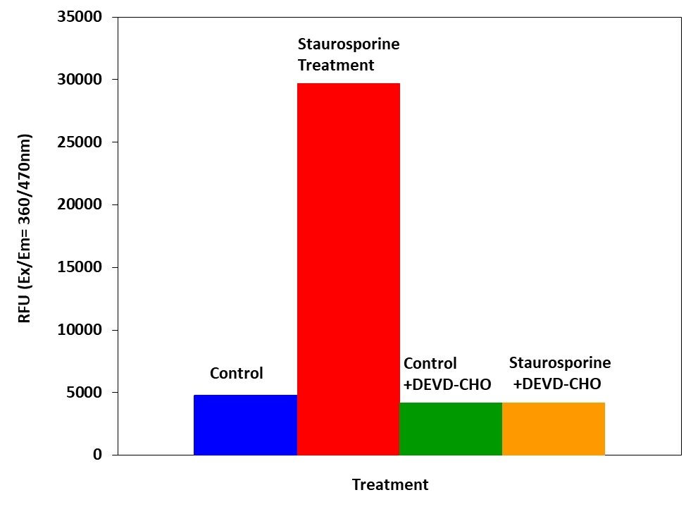
Detection of Caspase 3/7 activity in Jurkat cells with Cell Meter™ Caspase 3/7 Activity Apoptosis Assay Kit. Jurkat cells were seeded on the same day at 80,000 cells/well/90 µL in a Costar black wall/clear bottom 96-well plate. The cells were treated with or without 1 µM of staurosporine for 4 hours, and with or without 10 µM of the caspase inhibitor AC-DEVD-CHO for 10 minutes. The caspase 3/7 assay solution (100 µL/well) was added and incubated at room temperature for 1 hour. The fluorescence intensity was measured at Ex/Em = 360/470 nm (Cutoff = 420 nm).
Albumin exposure upregulates pro-apoptotic pathways in podocytes. A, Activated caspase 3/7 activity normalized to total cellular protein in podocytes treated with 5 mg/ml albumin (closed bars) or 5 mg/ml dextran (open bars) as an oncotic control for the indicated amounts of time. * denotes P&lt;0.0001 compared to corresponding dextran treated control. B, TUNEL staining of cultured podocytes treated with 5 mg/ml dextran (Dex) or 5 mg/ml albumin (Alb). Nuclei are stained blue with DAPI. TUNEL-positive nuclei are green. C, Caspase 3/7 activity normalized to total cellular protein in podocytes treated with 5 mg/ml albumin or 5 mg/ml albumin +50 &micro;M z-VAD, a pan-caspase inhibitor. z-VAD largely abrogated caspase activity (*, P = 0.01 versus albumin+z-VAD). D, Percentage of dead cells measured using the trypan blue assay in podocytes treated with 5 mg/ml albumin alone (open bar) or 5 mg/ml albumin +50 &micro;M z-VAD (closed bar) for 24 hrs or 5 mg/ml albumin alone (horizontal hatched bar) or 5 mg/ml albumin +50 &micro;M z-VAD for 48 hrs (vertical hatched bar). * denotes P = 0.001 compared to albumin+z-VAD at 48 hrs. *Caspase activity was determined using the Caspase 3/7 Activity kit (AAT Bioquest, Sunnyvale, CA) according to the manufacturer&rsquo;s directions. Source: Graph from <strong>Endocytosis of Albumin by Podocytes Elicits an Inflammatory Response and Induces Apoptotic Cell Death</strong> by Kayo Okamura, et al., <em>PLoS ONE</em>, Jan. 2013.