Cell Meter™ Caspase 9 Activity Apoptosis Assay Kit *Red Fluorescence*
Ordering information
Price | |
Catalog Number | |
Unit Size | |
Quantity |
Additional ordering information
Telephone | 1-800-990-8053 |
Fax | 1-800-609-2943 |
sales@aatbio.com | |
International | See distributors |
Bulk request | Inquire |
Custom size | Inquire |
Shipping | Standard overnight for United States, inquire for international |
Spectral properties
Excitation (nm) | 532 |
Emission (nm) | 619 |
Storage, safety and handling
H-phrase | H303, H313, H333 |
Hazard symbol | XN |
Intended use | Research Use Only (RUO) |
R-phrase | R20, R21, R22 |
UNSPSC | 12352200 |
Alternative formats
Cell Meter™ Caspase 9 Activity Apoptosis Assay Kit *Blue Fluorescence* |
Cell Meter™ Caspase 9 Activity Apoptosis Assay Kit *Green Fluorescence* |
Related products
Overview | ![]() ![]() |
See also: Caspases
Excitation (nm) 532 | Emission (nm) 619 |
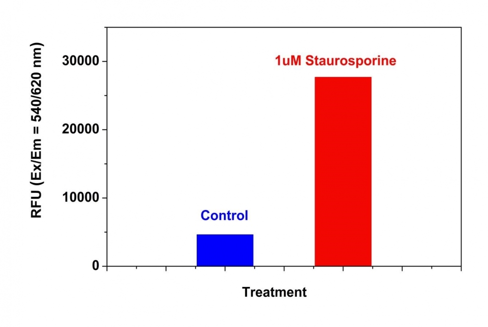
Our Cell Meter™ assay kits are a set of tools for monitoring cellular functions. There are a variety of parameters that can be used. This particular kit is designed to monitor cell apoptosis by measuring caspase 9 activity. Caspase 9 is a member of the CED-3 subfamily. Activated Caspase-9 cleaves downstream caspases such as caspase-3, -6 and -7, initiating the caspase cascade. It is essential for apoptosis during normal development of the central nervous system. Caspase 9 is proven to have selectivity for the peptide sequence Leu-Glu-His-Asp (LEHD). This kit uses Ac-LEHD-ProRed™ as a fluorogenic indicator for caspase 9 activity. Cleavage of ProRed™ by caspase 9 generates strongly fluorescent ProRed™. The kit provides all the essential components. The assay is robust and can be readily adapted for high throughput screening. It can be used to either quantify the activated caspase 9 activities in apoptotic cells or screen the caspase 9 inhibitors. Quite a few labs have used this kit for high throughput screenings.
Platform
Fluorescence microplate reader
Excitation | 540 nm |
Emission | 620 nm |
Cutoff | 610 nm |
Recommended plate | Black wall/clear bottom |
Instrument specification(s) | Top or bottom read mode |
Components
Example protocol
AT A GLANCE
Protocol summary
- Prepare cells with test compounds (100 µL/well/96-well plate or 25 µL/well/384-well plate)
- Add equal volume of Caspase 9 Substrate working solution (100 µL/well/96-well plate or 25 µL/well/384-well plate)
- Incubate at room temperature for 1 hour
- Monitor fluorescence intensity (top or bottom read mode) at Ex/Em = 540/620 nm (Cutoff = 610 nm)
Important notes
Thaw all the kit components at room temperature before starting the experiment.
PREPARATION OF WORKING SOLUTION
Add 5 µL of 200X Ac-LEHD-ProRed™ stock solution (Component A) into 1 mL of Assay Buffer (Component B) and mix well to make Caspase 9 Substrate working solution. Protect from light. Note: Caspase 9 working solution is not stable, use it promptly. 1 mL of Caspase 9 Substrate working solution is enough for 10 assays. Note: Aliquot and store unused caspase 9 substrate (Component A) and assay buffer (Component B) at-20 oC. Avoid repeated freeze/thaw cycles.
For guidelines on cell sample preparation, please visit
https://www.aatbio.com/resources/guides/cell-sample-preparation.html
SAMPLE EXPERIMENTAL PROTOCOL
- Treat cells by adding 10 µL/well of 10X test compounds (96-well plate) or 5 µL/well of 5X test compounds (384- plate) into PBS or the desired buffer. For blank wells (medium without the cells), add the same amount of compound buffer.
- Incubate the cell plate in a 37 °C, 5% CO2, incubator for a desired period of time (3-4 hours for Jurkat cells treated with staurosporine) to induce apoptosis.
- Add 100 µL/well/96-wel or 25 µL/well/384-well plate of Caspase 9 Substrate working solution.
- Incubate the plate at room temperature for at least 1 hour, protected from light. Note: If desired, add 1 µL of the 1 mM Ac-LEHD-CHO caspase 9 inhibitor to selected samples 10 minutes before adding Caspase 9 Substarte working solution at room temperature to confirm the inhibition of the caspase 9-like activities.
- Monitor the fluorescence intensity with a fluorescence microplate reader (either top or bottom read mode) at Ex/Em = 540/620 nm (Cutoff = 610 nm). Note: Sometimes, bottom read gives better signal to background ratio. Centrifuge cell plate (especially for the non-adherent cells) at 800 rpm for 2 minutes (brake off) if using bottom read mode.
Product Family
Name | Excitation (nm) | Emission (nm) | Extinction coefficient (cm -1 M -1) |
Cell Meter™ Caspase 9 Activity Apoptosis Assay Kit *Blue Fluorescence* | 341 | 441 | - |
Cell Meter™ Caspase 9 Activity Apoptosis Assay Kit *Green Fluorescence* | 500 | 522 | 80000 |
Images

Figure 1. Detection of Caspase 9 Activities in Jurkat cells using Cell Meter™ Caspase 9 Activity Apoptosis Assay Kit *Red Fluorescence*. Jurkat cells were seeded on the same day at 300,000 cells/90 µL/well in a Costar black wall/clear bottom 96-well plate. The cells were treated with or without 1 µM of staurosporine for 4 hours. The caspase 9 Substrate working solution (100 µL/well) was added and incubated at room temperature for 1 hour. The fluorescence intensity was measured at Ex/Em = 540/620 nm (Cutoff = 610 nm) with FlexStation fluorescence microplate reader (Molecular Devices).
Citations
View all 16 citations: Citation Explorer
Contact-dependent signaling triggers tumor-like proliferation of CCM3 knockout endothelial cells in co-culture with wild-type cells
Authors: Rath, Matthias and Schwefel, Konrad and Malinverno, Matteo and Skowronek, Dariush and Leopoldi, Alexandra and Pilz, Robin A and Biedenweg, Doreen and Bekeschus, Sander and Penninger, Josef M and Dejana, Elisabetta and others,
Journal: Cellular and Molecular Life Sciences (2022): 1--20
Authors: Rath, Matthias and Schwefel, Konrad and Malinverno, Matteo and Skowronek, Dariush and Leopoldi, Alexandra and Pilz, Robin A and Biedenweg, Doreen and Bekeschus, Sander and Penninger, Josef M and Dejana, Elisabetta and others,
Journal: Cellular and Molecular Life Sciences (2022): 1--20
Combination of Goniothalamin and Sol-Gel-Derived Bioactive Glass 45S5 Enhances Growth Inhibitory Activity via Apoptosis Induction and Cell Cycle Arrest in Breast Cancer Cells MCF-7
Authors: Bakar, Siti Aishah Abu and Ali, Abdul Manaf and Noor, Siti Noor Fazliah Mohd and Hamid, Shahrul Bariyah Sahul and Azhar, Nur Asna and Mohamad, Noor Muzamil and Ahmad, Nor Hazwani
Journal: BioMed Research International (2022)
Authors: Bakar, Siti Aishah Abu and Ali, Abdul Manaf and Noor, Siti Noor Fazliah Mohd and Hamid, Shahrul Bariyah Sahul and Azhar, Nur Asna and Mohamad, Noor Muzamil and Ahmad, Nor Hazwani
Journal: BioMed Research International (2022)
Promotion of osteointegration under diabetic conditions by tantalum coating-based surface modification on 3-dimensional printed porous titanium implants
Authors: Wang, Lin and Hu, Xiaofan and Ma, Xiangyu and Ma, Zhensheng and Zhang, Yang and Lu, Yizhao and Li, Xiang and Lei, Wei and Feng, Yafei
Journal: Colloids and Surfaces B: Biointerfaces (2016): 440--452
Authors: Wang, Lin and Hu, Xiaofan and Ma, Xiangyu and Ma, Zhensheng and Zhang, Yang and Lu, Yizhao and Li, Xiang and Lei, Wei and Feng, Yafei
Journal: Colloids and Surfaces B: Biointerfaces (2016): 440--452
microRNA-186 inhibits cell proliferation and induces apoptosis in human esophageal squamous cell carcinoma by targeting SKP2
Authors: He, Wei and Feng, Jianfang and Zhang, Yan and Wang, Yuanyuan and Zang, Wenqiao and Zhao, Guoqiang
Journal: Laboratory Investigation (2016): 317--324
Authors: He, Wei and Feng, Jianfang and Zhang, Yan and Wang, Yuanyuan and Zang, Wenqiao and Zhao, Guoqiang
Journal: Laboratory Investigation (2016): 317--324
Hypoxia regulates TRAIL sensitivity of colorectal cancer cells through mitochondrial autophagy.
Authors: Knoll, Gertrud and Bittner, Sebastian and Kurz, Maria and Jantsch, Jonathan and Ehrenschwender, Martin
Journal: Oncotarget (2016)
Authors: Knoll, Gertrud and Bittner, Sebastian and Kurz, Maria and Jantsch, Jonathan and Ehrenschwender, Martin
Journal: Oncotarget (2016)
Role of delta-like ligand-4 in chemoresistance against docetaxel in MCF-7 cells
Authors: Wang, Q and Shi, Y and Butler, HJ and Xue, J and Wang, G and Duan, P and Zheng, H
Journal: Human & Experimental Toxicology (2016): 0960327116650006
Authors: Wang, Q and Shi, Y and Butler, HJ and Xue, J and Wang, G and Duan, P and Zheng, H
Journal: Human & Experimental Toxicology (2016): 0960327116650006
Angiopoietins Modulate Survival, Migration, and the Components of the Ang-Tie2 Pathway of Chronic Lymphocytic Leukaemia (CLL) Cells In Vitro
Authors: Palma, Luis Mario Aguirre and Flamme, Hanna and Gerke, Iris and Kreuzer, Karl-Anton
Journal: Cancer Microenvironment (2016): 13--26
Authors: Palma, Luis Mario Aguirre and Flamme, Hanna and Gerke, Iris and Kreuzer, Karl-Anton
Journal: Cancer Microenvironment (2016): 13--26
t-BHQ Provides Protection against Lead Neurotoxicity via Nrf2/HO-1 Pathway
Authors: Ye, Fang and Li, Xiaoyi and Li, Lili and Yuan, Jing and Chen, Jun
Journal: Oxidative medicine and cellular longevity (2015)
Authors: Ye, Fang and Li, Xiaoyi and Li, Lili and Yuan, Jing and Chen, Jun
Journal: Oxidative medicine and cellular longevity (2015)
Insulin improves osteogenesis of titanium implants under diabetic conditions by inhibiting reactive oxygen species overproduction via the PI3K-Akt pathway
Authors: Wang, Lin and Zhao, Xiong and Wei, Bo-yuan and Liu, Yi and Ma, Xiang-yu and Wang, Jian and Cao, Peng-chong and Zhang, Yang and Yan, Ya-bo and Lei, Wei and others, undefined
Journal: Biochimie (2015): 85--93
Authors: Wang, Lin and Zhao, Xiong and Wei, Bo-yuan and Liu, Yi and Ma, Xiang-yu and Wang, Jian and Cao, Peng-chong and Zhang, Yang and Yan, Ya-bo and Lei, Wei and others, undefined
Journal: Biochimie (2015): 85--93
Glucose promotes cell proliferation, glucose uptake and invasion in endometrial cancer cells via AMPK/mTOR/S6 and MAPK signaling
Authors: Han, Jianjun and Zhang, Lu and Guo, Hui and Wysham, Weiya Z and Roque, Dario R and Willson, Adam K and Sheng, Xiugui and Zhou, Chunxiao and Bae-Jump, Victoria L
Journal: Gynecologic oncology (2015): 668--675
Authors: Han, Jianjun and Zhang, Lu and Guo, Hui and Wysham, Weiya Z and Roque, Dario R and Willson, Adam K and Sheng, Xiugui and Zhou, Chunxiao and Bae-Jump, Victoria L
Journal: Gynecologic oncology (2015): 668--675
Application notes
FAQ
Are inflammasomes and caspase-1 related?
Do you offer any fluorimetric assays that measure caspase activation/activity in live cells using a flow cytometer?
Does pH and staining temperature affect Annexin V-Phosphatidylserine binding?
Does propidium iodide stain apoptotic cells?
How can I tell if my cell sample is dying?
Do you offer any fluorimetric assays that measure caspase activation/activity in live cells using a flow cytometer?
Does pH and staining temperature affect Annexin V-Phosphatidylserine binding?
Does propidium iodide stain apoptotic cells?
How can I tell if my cell sample is dying?