Products
Services
Resources
Selection Guides
About
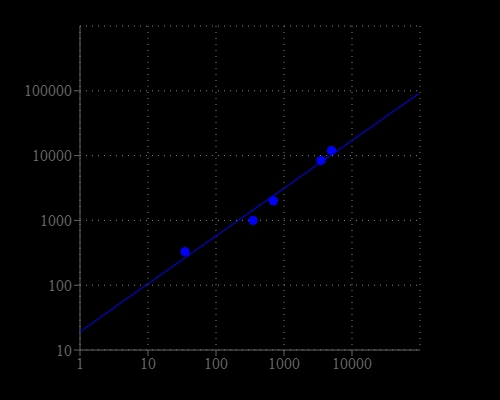
Cell Meter™ Cell Viability Assay Kit
Blue Fluorescence with 405 nm Excitation
There are a variety of parameters that can be used to monitor cell viability. The proprietary violet laser -excitable fluorescent dye used in the kit is a hydrophobic compound that easily permeates intact live cells and gets enhanced fluorescence upon entering into live cells. The hydrolysis of the non-fluorescent substrate by intracellular esterases generates a strongly blue fluorescent product that is well-retained in the cell cytoplasm. The blue fluorophore generated by the esterase hydrolysis of the non-fluorescent substrate has the spectral properties of fluorescein. When excited at 405 nm, the fluoreophore emits intense blue fluorescence at ~450 nm. The kit provides all the essential components with an optimized cell-labeling protocol for fluorescence microplate assays. This Cell Meter™ Cell Viability Assay Kit provides an effective tool of labeling cells for fluorescence flow cytometry, microplate and microscopic investigations of cellular functions. It is useful for a variety of studies, including cell adhesion, chemotaxis, multidrug resistance, cell viability, apoptosis and cytotoxicity. The kit is suitable for proliferating and non-proliferating cells.
| Catalog | Size | Price | Quantity |
|---|---|---|---|
| 22784 | 500 Tests | Price |

Spectral properties
| Excitation (nm) | 406 |
| Emission (nm) | 445 |
Storage, safety and handling
| H-phrase | H303, H313, H333 |
| Hazard symbol | XN |
| Intended use | Research Use Only (RUO) |
| R-phrase | R20, R21, R22 |
| UNSPSC | 12352200 |
Instrument settings
| Fluorescence microplate reader | |
| Excitation | 405 nm |
| Emission | 460 nm |
| Cutoff | 435 nm |
| Recommended plate | Black wall/clear bottom |
| Instrument specification(s) | Bottom read mode |
Contact us
| Telephone | |
| Fax | |
| sales@aatbio.com | |
| International | See distributors |
| Bulk request | Inquire |
| Custom size | Inquire |
| Technical Support | Contact us |
| Request quotation | Request |
| Purchase order | Send to sales@aatbio.com |
| Shipping | Standard overnight for United States, inquire for international |
Page updated on June 17, 2026